Gastrointestinal disorders in the
cavalier King Charles spaniel
-
 The
Gut - What It Is
 - List of Disorders
- Fecal Scoring
- Diagnosis
- Treatment
- Research News
- Related Links
- Veterinary Resources
Cavalier King Charles spaniels have been found to be among the five breeds with the highest prevalence for gastrointestinal disease. Gastrointestinal diseases in CKCSs are varied and include chronic and acute ones. They have varied causes, and they have varied consequences to the cavaliers' health. They include: chronic enteropathy (CE), inflammatory bowel disease (IBD), hemorrhagic gastroenteritis (HGE), hernia, protein-losing enteropathy (PLE).
In a 2015 review of the prevalence of disorders in 1,875 cavaliers in England in the VetCompass database, gastrointestinal disorders were the fourth most common (behind heart, skin, and eyes), affecting 19.3% of all CKCSs in the study. Broken down by category, gastrointestinal included diarrhea, gastritis, colitis, eneritis, gastroenteritis, enterocolitis, gastrointestinal infections, foreign bodies, constipation, IBD, fecal incontinence, intestinal masses, rectal prolapse, PLE, and megaesaphagus. In a 2015 review of breed pattern of diagnosis in Japan in 2010, including 5,743 cavaliers, they had a prevalance of 21% for digestive system disorders.
The Gut -- What It Is
The gastrointestinal system is the dog's digestive tract (also, the gut. GI tract, alimentary canal), starting with the mouth, then the esophagus, the stomach, intestines (small and large -- colon or bowel), rectum, and anus. It is the means of transfering food to energy and nutrients and expelling its waste. In general, the GI tract refers to just the stomach and intestines.
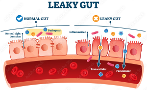
The intestinal walls -- intestinal barrier -- consist of three layers (see image at right):
(1) the mucosal (mucus) layer, composed of water, glycoproteins, and antimicrobial proteins;
(2) the epithelial layer, which are porous cell membranes in the sense that they allow certain items to pass through to the blood stream, and they prevent others, particularly toxins and bacteria, from entering the blood stream; and
(3) the lamina propria, which contains immune cells such as plasma cells and macrophages.
There also is a direct connection between the gastrointestinal system and the liver. For instance, the portal vein transports nutrient-rich blood from the GI tract to the liver for further processiung and conversion to energy. These nutrients include proteins, carbohydrates, and fats for processing, along with toxins and waste products to be detoxified by the liver.
RETURN TO TOP
List of Disorders
Here we list alphabetically and discuss in some detail those gastrointestinal diseases found to be most prevalent in the CKCS, according to specific published studies.
- Angiodysplasia
- Chronic enteropathy
- Colitis (colonitis)
- Diarrhea (Diarrhoea)
- Dysbiosis
- Esophagus disorders
- Gastroesophageal reflux disease (GERD)
- Megaesophagus
- Acute hemorrhagic diarrhea syndrome (AHDS)
- Hernia
- Inflammatory bowel disease (IBD)
- Leaky gut syndrome
- Polyp
- Protein-losing enteropathy (PLE)
- Related disorders
RETURN TO TOP
Angiodysplasia
Angiodysplasia (AGD -- also called vascular ectasia) is a fragile
malformation of a blood vessel -- artery, vein, or
 capillary
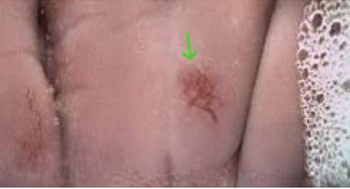
-- which can appear in any segment of the GI tract. If and when an AGD
is disrupted, a lesion develops which appears as bright red and flat and
consists of a cluster of dilated capillaries within the mucosal layer of
the GI tract. (See the green arrow in the photo.) Although relatively
rare, it has been detected using video capsule endoscopy (VCE) in a
cavalier among 15 dogs out of 291 in
this March 2023 article.
RETURN TO TOP
Chronic enteropathy
Any gastro-intestinal disease which lasts at least 3 weeks is definied as chronic enteropathy (CE), when external causes, such as parasites or tumors, are ruled out. Such diseases include those listed elsewhere on this webpage. The definition of CE is based upon how long the symptoms have existed. Cavalier King Charles spaniels are included among breeds found to have an increased risk of CE. (See this June 2022 article.)
There are four categories of chronic enteropathy, all based upon their response to treatment:
(1) Food-responsive enteropathy (FRE)
(2) antibiotic-responsive enteropathy (ARE)
(3) immunosuppressant-responsive enteropathy (IRE) or steroid-responsive enteropathy
(4) non-responsive enteropathy (NRE).
Symptoms over the 3+ week period include persistent diarrhea,
vomiting, loss of appetite, and/or weight loss.
 Chronic vomiting and
diarrhea are the most common clinical signs of CE in dogs. See
this December 2019 article for more details.
Suspected causes vary but include a combination of the dog's genetics, external environmental factors, environment within the intestines, and the dog's immune system.
In a February 2023 article, a team of Finnish veterinary researchers drew up a questionnaire for Finland dog owners to complete, pertaining to types of foods fed while their dogs were puppies and the onset of chronic enteropathy later in their dogs' lives. Owners of 1,016 young puppies (aged 2 to 6 months) and of 699 adolesent puppies (aged 6 to 18 months) reported the subsequent onset of chronic enteropathy (CE) symptoms, amounting to 21.7% of young puppies and 17.8% of adolescents. Reported dogs which did not develop CE were 3,665 young puppies and 3,227 adolscents. In this study, the definition of CE included inflammatory bowel disease (IBD), chronic gastrointestinal symptoms and/or food 'allergies' resulting in chronic gastrointestinal symptoms. Food recipes were defined as RC1 (non-processed meat-based diet -- raw red meat, organ meats, fish, eggs, tripe, bones and cartilage, vegetables, berries and fruits; RC2 (home cooked diet of grains, vegetables, fish, egg, organ meats, but apparently not red meats); RC3 human food leftovers and table scraps (cooked potato, non-sour milk products, cooked poultry and fish, processed meat, such as sausages, cooked rice and grain products, blood pancakes, and liver casserole); UPCD (ulta-processed dry dog food). They report finding that:
"We found that feeding a non-processed meat-based diet and giving the dog human meal leftovers and table scraps during puppyhood (2-6 months) and adolescence (6-18 months) were protective against CE later in life. Especially raw bones and cartilage as well as leftovers and table scraps during puppyhood and adolescence, and berries during puppyhood were associated with less CE. In contrast, feeding an ultra-processed carbohydrate-based diet, namely dry dog food or 'kibble' during puppyhood and adolescence, and rawhides during puppyhood were significant risk factors for CE later in life. ... A home-cooked diet was not significantly associated with CE incidence later in life in this study."
In an April 2025 article, 20 dogs diagnosed with chronic enteropathy and not responding favoraly to dietary treatment, were treated with fresh (same day) processed feces which was inserted via enema. The dosage was 2.5 to 5 g feces per kg/body weight. The dogs were tested monthly for 90 days and then followed up long term for a year. They report that 17 of the 20 dogs clinically improved up to 90 days and 10 dogs remained clinically stable up to one year after FMT. For more inforamtion about FMT, see this section below.
Treatment trials are the main means of differentiating between the above categories of CE. A change of diet usually is the first step in diagnosing CE, and in most cases is successful. If not, antibiotics are the second step, followed by immunosuppressive corticorsteroid (e.g., prednisolone and/or methylprednisolone) trials if necessary. Also, gastroprotectant treatment with either proton pump inhibitors, sucralfate, or famotidine (Pepcid, Apo-Famotidine) may be administered. In cases where none of these treatments are successful, the clinician may perform an endoscopy, especially if the dog's symptoms are severe or such factors as hypoalbuminaemia (low level of albumin) are found in the bloodwork.
In a June 2022 article, Swedish researchers reviewed the medical records of 814 dogs, including 31 cavaliers (3.8%), diagnosed with chronic enteropathy at two Swedish referral hospitals. They describe CE as being characterized by persistent (symptoms longer than 3 weeks) or recurring gastrointestinal signs, such as diarrhea, vomiting, loss of appetite, and weight loss. They report finding that breeds with the highest relative risk of acquiring CE included, in order, Norwegian Lundehunds, West Highland white terriers, miniature poodles, border terriers, Rottweiler, boxers, cavaliers, French bulldogs, and Shetland sheepdogs.
RETURN TO TOP
Colitis
Colitis (colonitis) is a form of inflammatory bowel disease (IBD) which affects the colon, the large intestine. See that section below.
RETURN TO TOP
Diarrhea (Diarrhoea)
Canine acute diarrhea (diarrhoea) describes loose or liquid stools and increased frequency of evacuating the bowels. It is very common and in most cases requires no treatment -- or perhaps just fluids to avoid dehydration -- as it tends to cure itself, in the sense that within 3 to 7 days following its onset, it clears up. In cases which last longer, they are captioned as "prolonged" or "chronic" rather than acute.
The causes vary, and include reactions to changes in diet, consuming inappropriate foods, parasites, and mild bacterial infections. Diarrhea of unspecified cause was second only to mitral valve disease as the most common disorder diagnosed among cavalier King Charles spaniels, in the April 2015 UK study of 3,624 cavaliers treated at primary care veterinary practices.
As mild and as self-limiting as diarrhea may be, it is not to be ignored if it does not resolve itself within a week, because it can be a sign of more serious gastric disorders and also chronic pancreatis or exocrine pancreatic insufficiency (EPI).
Acute diarrhea is to be distinguished from a severe disorder also common among cavaliers, acute hemorrhagic diarrhea syndrome (AHDS) also known as hemorrhagic gastroenteritis (HGE), which includes bloody stools and is discussed below.
RETURN TO TOP
Dysbiosis
Dysbiosis is the loss of favorable bacteria in the gut microbiome, due to disruption of the intestinal barrier. This loss can result in the microbiome becoming unbalanced and disrupted, due to a lack of diversity in the types of microbes in dog's gut, or a deficiency of beneficial gut microbes , or an excess of harmful bacteria. Possible causes include infectious illnesses, such as periodontal disease, certain diets, or prolonged use of bacteria-destroying medications (antibiotics). Dysbiosis has been associated with inflammatory bowel disease and disorders elsewhere in the dog's body, including the central nervous system, canine cognitive dysfunction (CCD), and neuroinflammation (the gut-brain axis). See this February 2025 article for details.
RETURN TO TOP
Esophagus disorders
• Gastroesophageal reflux disease (GERD)
The esophagus is the muscle-lined tube which transports food from the mouth to the stomach. The more common disorder of the esophagus in dogs, including cavaliers, is gastroesophageal reflux disease (GERD). With GERD, the stomach contents leak back (reflux) into the esophagus and cause irritation to its musular lining. This is due to the failure of one of the esophagus' muscles to close the connection with the stomach.
In a November 2012 study, a team of Canadian researchers studied seven fly-biting dogs, including two cavaliers, and found that they were suffering from gastrointestinal disorders, including GERD. In this study, the researchers treated the gastrointestinal (GI) diseases and observed complete resolution of the fly-biting in five (including a cavalier) of six of the seven dogs. The seventh dog (the other cavalier) was diagnosed with Chiari-like malformation and responded temporarily to pain management. The researchers concluded that:
"Fly biting behaviour may be caused by an underlying medical disorder, GI disease being the most common. Resolution of this behaviour is possible following specific treatment of the underlying medical condition."
• Megaesophagus
Megaesophagus is a relatively rare disorder in which the muscles of the dog's esophagus lose their motion, with the result that the esophagus widens, and the dog is unable to swallow food. It usually will regurgitate the food.
There can be various causes of megaesophagus, most of which are idiopathic and fewer being secondary to other, underlying disorders. The most common cause of secondary megaesophagus is myasthenia gravis, an autoimmune disease of neuromuscular transmission. See this April 1996 article, in which a cavalier was one of fifteen canine patients diagnosed with megaesophagus due to focal myasthenia gravis.
RETURN TO TOP
Acute hemorrhagic diarrhea syndrome (AHDS)
Cavaliers appear to be more likely to develop acute hemorrhagic diarrhea syndrome (AHDS) * than the average purebred breed. It's cause is not known, but its symptoms are well noted -- primarily vomiting and bright red bloody diarrhea, appearing suddenly and without advance signs. The blood source is attributed to necrosis of the dog's intestinal mucosal lining. Other symptoms may include a decreased appetite, fever, fatigue, and a painful abdomen.
*Formerly known as hemorrhagic gastroenteritis (HGE).
Suspected causes include any of the following, singly or combined: pancreatitis, intestinal bacteria or parasites, infections, intestinal ulcers or tumors, canine parvovirus, stress, and/or anxiety.
It is very important that AHDS be diagnosed and treated immediately, to avoid dehydration and possible death. Diagnosis includes the process of eliminating other possible causes, and therefore requires a complete blood count and analysis, urinalysis, x-rays, fecal evaluation, and ultrasound or endoscopic examinations of the gastrointestinal tract.
Treatment includes intravenous fluid (IV) therapy, diet restriction, antibiotics, and intestinal medications. Dogs who suffer from AHDS once may be more likely to develop it in the future. In an April 2015 article, gastroenteritis was reported in only 11 cavaliers among 3,624 CKCSs treated at primary-care veterinary practices in England from 2009 to 2013. However, diarrhea, due to unspecified causes, was the second most common disorder in that report, with 193 cases, second only to heart murmurs. Also, clinical reports of diagnosing and treating cavaliers with AHDS are abundant.
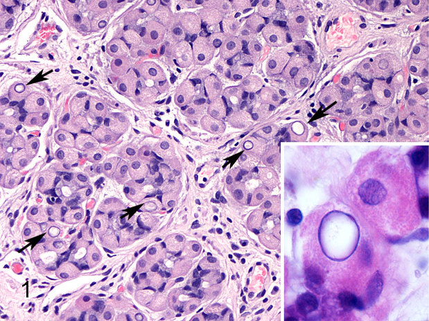 In
a January 2017 article, a team of Italian veterinary pathologists
report that cavaliers were the main represented
purebred breed in their study of nuclear glycogen inclusions in canine
parietal cells -- cells which are located in the gastric glands found in
the lining of the fundus and in the body of the stomach. Mixed-breed
dogs were 26% (5 dogs) of the dogs in the 24-dog study found to have
these inclusions, and cavaliers were second with 11% (2 dogs).
(Images at right: Nested gastric glands containing nuclear inclusions in
parietal cells [arrows]. Inset: detail of a nuclear inclusion in a
parietal cell.)
They concluded:
"Our findings suggest that nuclear glycogen inclusions in canine parietal cells could be an incidental finding. Nevertheless, since nuclear glycogen is present in several pathologic conditions, further investigations could be warranted to determine their true significance."
At this point, they reported that it is not known that findings of nuclear glycogen inclusions in the cavaliers' stomach glands are related to AHDS.
In a February 2021 article, a team of University of Copenhagen (Denmark) researchers examined the medical records of 237 dogs hospitalized for suspected AHDS. Twelve (5%) of the dogs were cavaliers, ranking the breed fourth behind only Labrador retrievers, small mixed breeds, and miniature Schnauzers, out of a total of 70 breeds overall. The researchers reviewed the disease's severity using:
• AHDS index: the degrees of severity -- normal, mild, moderate, or severe -- of six categories -- activity, appetite, stool consistency, stool frequency, vomiting frequency, and dehydration %);
• Systemic inflammatory response syndrome (SIRS) criteria: Hypo- or hyperthermia, tachycardia (heart rate in beats per minute), tachypnea (respiratory rate in breaths per minute), leukipenia or leukocytois (# of cells), immature band neutrphils (3 of cells), and hypoglycemia;
• Serum C-reactive protein (CRP) levels upon admission. They report that the majority of hospitalized dogs improved rapidly with only symptomatic treatment, meaning not including antimicrobials.
They found that the SIRS criteria may be a "poor proxy" for identifying affected dogs needing antimicrobial treatment, and that the role of CRP needs further investigation.
RETURN TO TOP
Hernia
Two cases of pleuroperitoneal diaphragmatic hernia have been reported in a total of six cavalier King Charles spaniels.
 In a
November 2019 article, UK and Swiss researchers reported on five separate cases of
cavalier King Charles spaniels diagnosed with a congential defect in the
diaphram causing the stomach and/or other abdominal organs to herniate
into the chest cavity. All of the affected cavaliers were intact males,
four of the
between 2.5 months and 9 months old, and the fifth dog was 18 months
old. The cases occurred over a period of 12 years. The symptoms were
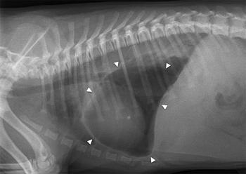
rapid breathing and shortness of breath. In all of the dogs, chest
x-rays showed a gas-fluid-filled structure on the left side of the
chest, which was found to be the stomach (see photo at right), with some degree of lung collapse and shift to the opposite side.
The clinicians determined that the dogs developed "tension
gastrothorax", which describes the stomach herniating through a
congenital diaphragmatic defect into the thorax and distended due to
being filled with trapped air. One of the CKCSs was euthanized, and
surgery was performed on the other four. The stomach was herniated into
the chest cavity in all five cases, and in others, the spleen and/or
omentum and/or intestine also was herniated. The hernias were repaired
and all four of the surviving dogs recovered successfully. The authors
concluded:
"In this report, all cases were purebred CKCS; they were presented in a timespan of approximately 12 years, and no information was available regarding their pedigree. Genetically inherited disease in CKCS includes mitral valve disease, syringomyelia, congenital and juvenile cataract and multi-focal retinal dysplasia. Based on this report, CKCS could also be a breed predisposed to pleuroperitoneal diaphragmatic hernia, but extensive breeding studies would be necessary to evaluate this hypothesis. Interestingly, all the cases were male, suggesting the possibility of a sex chromosomal mode of inheritance."
In an April 2021 article, Australian clinicians reported an 8-week-old cavalier puppy which had developed a congenital pleuroperitoneal hernia during an airplane flight from the breeder's home to the new owners' home. The onset of the hernia was attributed to the airplane's cabin pressure, which would have been no lower than 565 mmHg, the equivalent to an altitude of 8000 feet when the aircraft is at maximum operating altitude of 30,000 feet. According to Boyle's law, air in the gastrointestinal tract expands by approximately 30% as the atmospheric pressure is reduced during the flight, leading to increased intraabdominal pressure. The increased intra-abdominal pressure likely forced the abdominal viscera to move through the defect in the diaphragm. The intestinal distension in the enclosed thoracic space, likely lead to obstruction of the small intestines, resulting in significant compression of the already compromised lungs. Surgery was performed to relieve the conditions. The clinicians concluded:
"An acute presentation of respiratory distress after a high-altitude flight should alert clinicians to the possibility of sudden manifestation of a pleuroperitoneal hernia. ... As the condition of pleuroperitoneal hernia may have a genetic basis, counselling of owners and breeders in this regard is warranted."
RETURN TO TOP
Inflammatory bowel disease (IBD)
Inflammatory bowel disease (IBD) describes a group of gastrointestinal diseases which result in inflammation of the intestines and chronic symptoms related to gastrointestinal system. Colitis (colonitis), which is inflammation of the colon, is within that group. Hypersensitive immune system responses initiated by bacteria in the intestines is believed to be the cause of inflammation.
Symptoms of IBD usually include: diarrhea, painful bowel movements, mucous or blood in the stools, weight loss, chronic flatulence, vomiting, and gastric distention.
Several published studies have found that the cavalier King Charles spaniel experiences more than its fair share of these disorders. See these studies: September 1980, October 1995, December 2002, December 2008, May 2010, August 2011, March 2012, January 2013. Typical symptoms include primarily diarrhea, along with fatigue, vomiting, rumbling and gurgling abdominal sounds, and bright red blood in stools.
In the December 2002 article, the cavalier was diagnosed with eosinophilic enteritis, an uncommon form of IBD. Clinical signs were similar to other forms of IBD, but gastrointestinal ulceration and blood loss was detected, resulting in borderline anaemia.
In a December 2016 article, a team of University of California-Davis reasearchers examined the medical records from 1995 to 2010 of 90,090 dogs of all AKC-recognized breeds treated at their veterinary hospital, to determine the relation between neuter status and autoimmune diseases. They report finding that neutered dogs had a significantly greater risk of IBD.
In a May 2021 press release, FSD Pharma, Inc., a Canadian company, announced that it had applied to the US Food & Drug Administration (FDA) for approval to use FSD201 (ultramicronized palmitoylethanolamide, or ultramicronized PEA) to treat gastrointestinal enteropathy in dogs. The application reportedly has been accepted for review. The proposed trial design is a randomized, double-blind, placebo-controlled, crossover, trial comparing FSD201 dosed twice daily for 30 days to placebo for the treatment of canine inflammatory bowel disease. The primary endpoint will be a validated diarrhea score, evaluated by both treating veterinarian and dog owner. The trial will be conducted at 5-10 sites in the USA, and will enroll up to 200 dogs.
RETURN TO TOP
Leaky gut syndrome
 A
nickname for the dog's gastrointenstinal system is the "gut".
"Leaky" refers to excessive permeability of the intestinal walls,
particularly of the bowels. The intestinal walls consist of a layer of
mucus (mucosal layer) and a layer of
epithelial cells, which combined serve as the intestinal barrier IB). These cell membranes
are porous in the sense that they allow certain items to pass through to
the blood stream, and they prevent others, particularly toxins and
bacteria, from entering the blood stream.
Inflammation in the intestines damage the mucosal barrier inside the walls, allowing toxins and even undigested foods within to pass through the walls and into the bloodstream. This triggers more inflammation. Increased intestinal permeability (hyperpermeability) is concurrent with, and may be a cause of, immune system responses and liver dysfunction. The degree of intestinal permeability is measured by urinary testing, preferrably obtained with a urinary catheter following feeding long chain sugars which collect in the urine. Another test is to collect the dog's saliva to measure IgA and IgM. See this May 2025 article for details about the tests and nutitional treatments for leaky gut syndrome.
RETURN TO TOP
Polyp
Polyps in the epithelium lining of the stomach or intestinal wall are growths (lesions). In a September 2024 article, a 5-year-old cavalier experienced black tarry feces (melena), suggesting intestinal bleeding, and was vomiting blood (haematemesis) as well. Initially, she was treated with intravenous fluids and medications, and her signs improved. Abdominal ultrasound showed a mass in her duodendum. Gastrointestinal endoscopy identified a lesion in the duodendum, and biopsies indicated an epithelial duodenal polyp, meaning a growth in the lining of the upper small intestine. The clinicans surgically removed the portion of the duodendum where the polyp was located and resectioned the duodendum successfully. They determined that the polyp was hyperplastic, meaning non-cancerous. A year later, the dog experienced no polyp regrowth or clinical signs.
RETURN TO TOP
Protein-losing enteropathy
 Protein-losing enteropathy
(PLE) is the excess loss of proteins (albumin and globulin) in the intestines, caused
by any of a variety of diseases, including inflammation (iPLE), infiltration,
ulceration, blood loss, and particularly,
lymphangiectasia (dilation of lymph
vessels). Low serum protein in blood tests is the means of
detecting this disorder.
Yorkshire terrier, soft-coated Wheaton terrier, Norwegian Lundehund, and Basenji reportedly are predisposed to PLE, but there are reported cases of cavaliers being diagnosed with the syndrome. See these January 2011, March 2011, September 2013, and May 2015 articles.
Because there can be any of several different causes of PLE, the first step always is to determine the cause and treat that cause. The initial symptom of PLE usually is loss of weight due to malnutrition, but other signs may include intermittent diarrhea and vomiting. Since any of a number of disorders may be the cause of the PLE, an initial goal is to determine whether or not gastro-intestinal (GI) tract is the origin of the protein loss. Once diagnosed, treatment options include dietary changes and management of the inflammation.
In an October 2022 article studying the rates of relapse of dogs following treatment for PLE, cavaliers were the second most prominent breed, behind Staffordshire bull terriers. The researchers found that inflammatory PLE is associated with a high rate of relapse in dogs, and that adherence to dietary recommendations wouldt help prevent subsequent relapse in dogs with iPLE that attain initial remission.
In a July 2023 article, UK researchers devised an undernutrition screening score (USS) for use at the time of diagnosis of protein-losing enteropathy (PLE) in dogs. The USS is calculated at the time of diagnosis and is based upon the presence and severity of five variables: appetite, weight loss, and body, muscle, and coat condition, with higher scores reflecting worse undernutrition. They report that the cavalier King Charles spaniel (8%) was most common breed among the 50 purebred patients and 7 mixed breeds. They found that dogs that failed to reach remission of their PLE symptoms within six months had higher USS scores at diagnosis compared with dogs that did reach remission. Of the five variables assessed in the USS, they concluded that a combination of epaxial muscle loss and coat condition was most predictive of not reaching clinical remission of PLE within six months.
In a May 2021 article, UK researchers reviewed the treatment of 57 dogs diagnosed with PLE. Cavaliers and cocker spaniels were the two breeds with the most dogs in the study, at five each (8.8%), of a total of 31 breeds plus crossbreeds. The study focused upon comparing survival outcomes from treatment with and without feeding tubes inserted directly into the stomach or small intestine (enteral feeding tubes). A positive outcome was defined as survival longer than 6 months or death due to unrelated causes. They report finding that, of the 21 dogs that had a feeding tube inserted within 5 days of diagnosis, 16 (76%) had a positive outcome and 5 (24%) had a negative outcome. Dogs treated with dietary treatment alone and dogs with an enteral feeding tube were significantly associated with a positive outcome. They concluded that enteral feeding in dogs with inflammatory PLE could be associated with improved treatment outcome, especially in those receiving immunosuppressive treatment, and should be considered in the treatment plan of these dogs.
Conventional treatment typically includes high protein and low fat diets. Homemade diets are frequently prescribed, to provide meals high in proteins (e.g., 80 to 85 grams per 1,000 kcal) and low in fats. Diets with as little as 7 grams of fat per 1,000 kcal have been prescribed in such cases. A diet with moderate fat for healthy dogs rarely is lower than 30 grams. Seafoods such as white fish or shrimp may be the main sources of proteins in such recipes.
Medium chain triglycerides (MCTs) may be recommended in addition to diets rich in proteins, but some cavaliers have been found to have a genetic mutation in which MCTs may cause seizures and other severe disorders. Including MCTs , such as coconut oil, is not adviseable unless the cavalier patient's DNA has been tested and found to not carry the mutation. See our MCT webpage for moer information about this disorder in our breed.
In a May 2021 press release, FSD Pharma, Inc., a Canadian company, announced that it had applied to the US Food & Drug Administration (FDA) for approval to use FSD201 (ultramicronized palmitoylethanolamide, or ultramicronized PEA) to treat gastrointestinal enteropathy in dogs. See our PEA webpage for more information about PEA in general. The application reportedly has been accepted for review. The proposed trial design is a randomized, double-blind, placebo-controlled, crossover, trial comparing FSD201 dosed twice daily for 30 days to placebo for the treatment of canine inflammatory bowel disease. The primary endpoint will be a validated diarrhea score, evaluated by both treating veterinarian and dog owner. The trial will be conducted at 5-10 sites in the USA, and will enroll up to 200 dogs.
RETURN TO TOP
Related disorders
• Fly biting
In a November 2012 study, a team of Canadian researchers studied seven fly-biting dogs, including two cavaliers, and found that they were suffering from gastrointestinal disorders, including eosinophilic and lymphoplasmacytic infiltration of the stomach and small bowel, delayed gastric emptying, and gastroesophageal reflux disease (GERD). In this study, the researchers treated the gastrointestinal (GI) diseases and observed complete resolution of the fly-biting in five (including a cavalier) of six of the seven dogs. The seventh dog (the other cavalier) was diagnosed with Chiari-like malformation and responded temporarily to pain management. The researchers concluded that:
"Fly biting behaviour may be caused by an underlying medical disorder, GI disease being the most common. Resolution of this behaviour is possible following specific treatment of the underlying medical condition."
See also our Fly Catchers Syndrome webpage.
• Excessive licking
In this July 2012 article, researchers concluded that GI disorders should be considered when diagnosing canine excessive licking of surfaces (ELS).
• Pancreatitis
Pancreatitis has been determined to be a cause of acute hemorrhagic diarrhea syndrome (AHDS). We have a separate webpage discussing pancreatitis.
• Anal sac disorders
Gastrointestinal disorders can be a cause of anal sac disorders, primarily as a result of the dog not producing feces adequate to squeeze secretions from the anal sacs as the dog defecates. We have a separate webpage discussing anal sac disorders.
• Mitral valve disease (MVD)
In a November 2021 article, Japanese researchers investigated the effects of mitral valve disease (MVD or MMVD), on the intestines in Chihuahuas. Based upon human research, they hypothesized that intestinal mucosal injury could be a complication of MVD in dogs and that the markers of intestinal mucosal injury would be significantly increased in dogs with MVD. ... Their main objective was to clarify the relationship between MVD and intestinal mucosal injury markers such as serum intestinal fatty acid-binding protein (I-FABP) and D/L-lactate concentrations. Sixty-nine Chihuahuas were divided into the four MVD stages (B1, B2, C, and D). They report finding that I-FABP was significantly higher in stage C/D Chihuahuas than in the two other groups, and stage B2 was significantly higher than healthy dogs; D-lactate was significantly increased in stages B2 and C/D compared to healthy dogs and stage B1. L-lactate was significantly higher in stage C/D Chihuahuas than in any other group, and L-lactate also was significantly higher in stage B2 than that in healthy dogs and stage. They concluded:
"This study showed that intestinal mucosal injury markers such as I-FABP and D-Lactate increased with the severity of MMVD in Chihuahuas and that Chihuahuas with MMVD who had heart failure were at particular risk for intestinal mucosal injury. This study is the first to investigate the relationship between heart disease and intestinal mucosa in Chihuahuas and demonstrate a potential relationship between MMVD morbidity and intestinal mucosal cell injury. ... In the future, if I-FABP is studied in dogs and a cutoff value can be established, it could become a new prognostic factor for heart failure in dogs."
RETURN TO TOP
Fecal Scoring
 The
appearance of the dog's feces (excrement) is one of the ways
veterinarians diagnose some of these gastrointestinal conditions. Since
dogs rarely defacate in the examining room, the veterinarian will ask
the owner to watch the dog defacate at home and describe what the result
looks like.
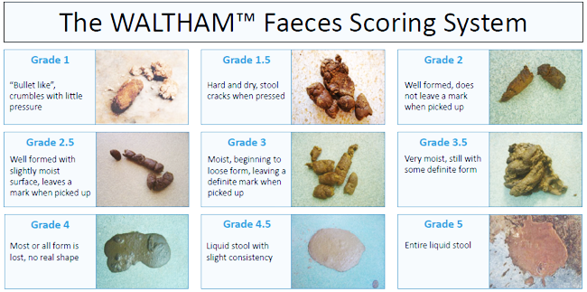
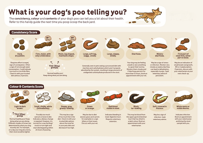
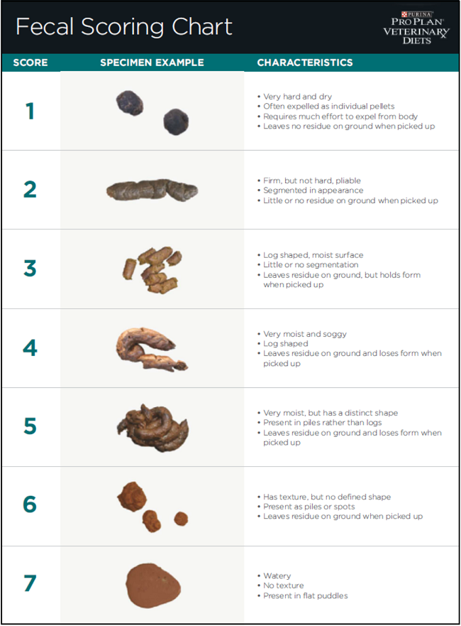
There are a few published ways to grade the quality of the resulting feces, as shown in the charts below. Being able to objectively categorize the dog's feces will be an aid to the veterinarian in attempting to diagnose and treat the disorder.
RETURN TO TOP
Diagnosis
In addition to visual fecal observation, laboratory examination of fecal samples include testing for infections, and DNA may be extracted from fresh samples for gene sequencing and analysis.
Food sensitivity testing may be performed to identify foods which may be associated with adverse effects in the GI system. An elimination diet may be performed for 2 to 3 months, removing any specific ingredient (protein, carbohydrate, fat) the dog has eaten within the past 6 months.
Microbiome tests, which measure the quantities and varieties of microbes in the GI system, will provide useful information in identifying any imbalances or deficiencies in the microbione. These tests involve extracting bacterial DNA from stool samples. This data will enable recipes to be drawn up which include ingredients to support the missing microbes. See this May 2024 article.
The canine microbial dysbiosis index (DI) is a patented method of identifying dysbiosis (the loss of favorable bacteria in the gut microbiome) in feces DNA by tracking the levels of various bacteria. The DI method enables clinicians to determine the difference between normal and abnormal bacteria health and whether the GI dysfunction is chronic (long term) or only acute (abbreviated). Chronic GI dysfunction may already have resulted in permanent changes to the patient's GI tract, requiring more of a management-based treatment than a curative one.
The DI uses a mathematical algorithm model based upon specific fecal bacteria quantities (total bacteria, Faecalibacterium, Turicibacter, Escherichia coli, Streptococcus, Blautia, Fusobacterium and Clostridium hiranonis) compared to a normal reference set. The model provides a single digit numerical value from a negative microbiota shift (below 0 -- normal or normobiosis) to mild microbiota shift to moderate shift to (2+) significant shift indicating dysbiosis. See this October 2017 article.
RETURN TO TOP
Treatment
Diet Changes
Diet changes may be advised and usually should be. In this September 2006 article, the authors stated:
"A 24-hour period of starvation, followed by the introduction of small frequent meals of bland food (eg, chicken and rice) is the most common treatment protocol for simple acute diarrhoea. Antibiotic therapy is not indicated unless a specific bacterial agent has been identified or there is evidence of intestinal ulceration (ie, haemorrhage)."
This fasting period may be followed by switching to a novel protein of hydrolyzed diet, and a low-fat diet, particularly if the diagnosis is IBD or PLE or lymphangiectasia.
This October 2023 article stated:
"Dietary indiscretion is incriminated in the majority of acute diarrhoea cases in dogs, with affected animals generally responding well to supportive treatment. Such treatment may include nutritional management, gastrointestinal nutraceutical and fluid therapy, with antimicrobials only recommended for dogs showing signs, or at high risk, of sepsis."
If the patient has been fed a dry (kibble) food, the way the dog consumes the kibble nuggets may have been inhibiting the digestive function, if the dog rarely actually chews and breaks down the nuggets before swallowing them. Switching to a wet food, preferrably home-prepared may support the digestion of the food and the absorption of nutrients.
Food that is cooked slowly at low temperatures provides the the GI system with the most easily digestible food and absorbable nutients. See this May 2024 article.
Fecal transplants, from canine donors with healthy GI microbiota, may be recommended. They are called fecal microbiome transplantation (FMT). Feces from dogs with healthful microbiota and a microbiome are given orally in capsules.
RETURN TO TOP
Medications
Antimicrobials: Considering the various types of gastro-intestinal diseases listed above, you may expect a variety of different forms of therapy. Metronidazole (Flagyl, Metizol, Protostat, Metrogel) typically is prescribed for treatment in the absence of a definitive diagnosis. It is an antimicrobial which may be used to treat inflammation in the large intestine, presumably due to bacterial infection and some parasites. However, metronidazole has not been found to be particularly successful in improving recovery. Metronidazole also has been found to cause long-term detrimental consequences for the gut microbiome and metabolome. See this July 2019 article and this October 2023 article.
Ironically, in an October 2022 article, researchers compared a 30-day treatment of noninfectious acute colitis in 59 dogs (none being cavaliers) with metronidazole in one group and with an "easily digestible diet" in a second group and the same diet plus powdered psyllium seed husk in a third group. They based the results upon the Waltham fecal score index. They found that the diet, with or without psyllium, was superior in managing the colitis compared to adding metronidazole. They stated: "The omission of metronidazole reduced the adverse impact significantly on intestinal microbiota."
H2-receptor antagonists: Famotidine (Pepcid, Apo-Famotidine) reduces stomach acid production, to treat inflammation, acid reflux, and GI ulcers. It may be given orally or by injection. It may not be appropriate for dogs also suffering heart, liver, and/or kidney disease or also being treated with certain other medications.
Lengthy immunosuppressive therapy with steroids may also be necessary.
RETURN TO TOP
Supplements
 If the
underlying causes are immune-related, treatment could include microbiome
support, such as supplementing probiotics and prebiotics, added
Vitamin B12 and folate (folic acid -- Vitamin B9).
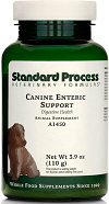
Standard Process offers its Canine Enteric Support as a supplement to suppor the dog's digestive system.
Powdered unflavored psyllium seed husk as a source of added fiber has been found successful in treating colitis, at the rate of a teaspoon to a tablespoon added to each meal. See this October 2022 article.
The Chinese herbal medicine, Da Xiang Lian Wan (DXLW), has been found to be an effective treatment for stress colitis in dogs. See this February 2017 article.
Palmitoylethanolamide (PEA): In a May 2021 press release, FSD Pharma, Inc., a Canadian company, announced that it had applied to the US Food & Drug Administration (FDA) for approval to use FSD201 (ultramicronized palmitoylethanolamide, or um-PEA) to treat gastrointestinal enteropathy in dogs. The application reportedly has been accepted for review. The proposed trial design is a randomized, double-blind, placebo-controlled, crossover, trial comparing FSD201 dosed twice daily for 30 days to placebo for the treatment of canine inflammatory bowel disease. The primary endpoint will be a validated diarrhea score, evaluated by both treating veterinarian and dog owner. The trial will be conducted at 5-10 sites in the USA, and will enroll up to 200 dogs.
Palmitoylethanolamide (PEA) is a
N-acylethanolamine molecule in a family of long-chain fatty acid
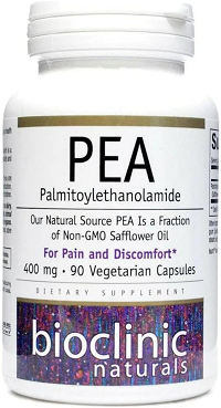 amides
called ALIAmides. PEA has been found in rat and mice studies to limit
hyperactvity in immune cells and thereby control inflammatory responses
and resulting tissue damage. PEA is produced by the animal's body as
needed in response to certain types of injuries. PEA is a product of
normal fatty acid synthesis from palmitic acid. It is found in many
common foods, particularly palm oil, soy beans, egg yolks, and peanuts.
The commercial version is most commonly manufactured from palm oil*.
Not all PEA is alike. There are at least 4 types of PEA, and the differences of those 4 are described below here. In short, since the basic (naïve) PEA is almost totally insoluble in water and therefore has very poor bioavailability, researchers use micronized or ultra-micronized PEA or water-dispersible PEA in their published studies.
• Basic PEA, called "naïve PEA", is almost totally insoluble in water and under gastrointestinal conditions and therefore the oral intake of it (rather than being injected directly into the abdomen) has very poor bioavailability, meaning that it does not get absorbed well in the dog's gut. See this May 2021 article and this July 2025 article.
• Micronized PEA (m-PEA or micro-palmitoylethanolamide) is a patented technique that reduces the diameter of PEA particles, making them absorbable in the intestine, which has been found to be more effective than ordinary basic PEA in activating PEA levels in blood plasma in dogs. See this August 2014 article.
• Ultra-micronized PEA (um-PEA), also patented, reduces the PEA particle size further, to enable it to cross the blood-brain barrier, likewise has been found to be much more effective than basic PEA. See this August 2014 article.
• Water-Dispersible PEA (PEA-WD), also patented, reduces the PEA to a powder which can be dispersed in cold water. It has been found to be 16 times more effective than basic PEA. See this July 2025 article.
• Hybrid versions of PEA: Additonally, PEA has been combined with other ingredients and used in some published studies. These include FenuMat-PEA (P-fen), which is a PEA hybrid combined with the herb fenugreek (trigonella foenum-graecum), and hybrids combined with resveratrol, quercetin, fisstin, and boswellic acid. See this July 2025 article.
PEA micronization and ultra-micronization are patented (by Italian company, EPITECH Group SpA) processing techniques that reduce the diameter of the PEA particle to a micronized or ultra-micronized size which optimizes the PEA's absorbability along the intestine. Micronization increases the drug's surface area, thereby improving its dissolution rate and minimizing its absorption difficulties. The ultra-micronized size also enables the PEA to cross the blood-brain barrier. See this February 2021 article.
 If a PEA product is not advertised as being micronized or
ultra-micronized, then
Dr. Clare Rusbridge advises that
"You
probably are wasting your money."
A variety of brands of
micronized and ultra-micronized PEA are offered on-line.
As for dosages, the studies using micronized PEA, the range was from 10 to 15 mg/kg/day, and the range for ultra-micronized was 24 mg/kg (for osteoarthritis in humans).
Read more about PEA on our PEA webpage.
* Palm oil: The palm oil cultivation industry has been destroying rainforests in Sumatra and Borneo in Indonesia and Malaysia, the only habitats of orangutans. If you are going to obtain PEA, we suggest that you do so only from vendors whose PEA has been manufactured with palm oil from sustainable sources and not the deforestation of rainforests. This link connects to a "PalmOil Scan Mobile App" which will enable you to determine if the PEA vendors you select obtain their palm oil from sustainable sources.
RETURN TO TOP
Fecal Microbiota Transplantation (FMT)
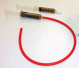 Fecal Microbiota Transplantation (FMT) is a process of stool
transplanting, whereby fecal material from a healthy donor dog is
inserted into the rectum of a dog suffering from an imbalance of
microbes in the gut, related to a variety of diseases. These diseases
primarily are directly associated with the gut, with most common
symptoms being diarrhea or vomiting and inflammation. A most common
disease is clostridium difficile infection (C-diff), an
antibiotic-resistant bacteria which can be fatal to dogs. The purpose of
this procedure is to supplement or replace the gut microbiota in the
dog's intestines, to increase the number and varieties of beneficial
bacteria to restore the proper function of the gut microbiota.
Microbiota is a combination of microorganisms (microbes) -- bacteria, fungi, protozoa, viruses, parasites -- which exist in the gut (stomach, intestines, rectum) of the host dog. The dog's microbiome is a combination of the genetic material in the microbes in the host. The microbiota in the intestines play important roles in maintaining the health of the dog, not just in the intestines but also in other organs, including the brain. A well balanced microbiome is essential for optimum well-being of the dog. When the microbiome becomes unbalanced, this disruption is called dysbiosis.
In 2020, a group of international veterinary specialists organized the Companion Animal FMT Consortium to develop guidelines and "best practices" for FMT in veterinary medicine. In 2024, this group published "Clinical Guidelines for Fecal Microbiota Transplantation in Companion Animals", to provide veterinarians with evidence-based recommendations for performing FMT in a variety of clinical practice settings. Among their guidelines are:
• Fecal collection: Ideally, naturally voided feces should be used, collected immediately after defecation.
• Fecal handling: Fresh feces should be processed and either administered to the patient or stored in an air-tight container as fast as possible and feasible (preferably within 2-6 hours of defecation).
• If immediate processing is not feasible, freshly voided feces should be stored refrigerated at 4ºC and processed as soon as possible.
• Processing should include removing large particles and foreign matter, creating a fecal slurry, and filtering using a strainer bag.
• When administered as a rectal enema, the consistency should be as thick as possible to keep the volume low and prevent leakage.
• If not used immediately, the fecal slurry should be frozen immediately and stored for future use, for optimal survival of the bacteria.
• Freeze drying fecal slurries to a powder form is available and being used at research and commercial facilities. The viability, shelf life, and clinical efficacy of freeze dried fecal powder in capsules is unknown.
• There is no evidence-based dosing regime for administering FMTs in any form (fresh, frozen, or freeze-dried) or through any route (rectal or oral).
The direct transplanting of the feces into the rectum of the patient dog is a medical procedure called endoscopy or colonoscopy or rectal enema, or it is given orally using a syringe, a feeding tube, or in a capsule. The Companion Animal FMT Consortium does not recommend administering FMT material through feeding tubes, and using an oral syringe may pose the risk of aspiration pneumonia. The lead author of the Companion Animal FMT Consortium's guidelines, Dr. Jenessa Winston, has stated, "Fresh is probably the best scenario. Anytime that we manipulate and freeze feces, we are going to have a change in viability."
In a November 2017 article about a human study, comparing the effectiveness of oral capsules and colonoscopy-delivered FMT to treat C-diff, the investigators reported that FMT via oral capsules was not inferior to delivery by colonoscopy. However, in a January 2024 article, in which 54 dogs were studied, fresh fecal material was given orally by capsule for 25 days. On average, only 18% of the stool donor's bacterial amplicon sequence variants (ASVs) engrafted in the FMT recipient. Since most capsules are designed to dissolve once they reach the stomach, and yet the goal of their FMT content is to not disperse until it reaches the intestines, there necessarily is a loss of effectiveness of the fecal material that is released before arriving at the intestines.
FMT currently is being used successfully to treat dogs for parvovirus and other causes of diarrhea and chronic enteropathy. At the time of publication of the Consortium's guidelines (2024), the authors report that the level of evidence or even anecdotal reports of using FMT for diseases apart from GI issues, such as diabetes mellitus and obesity, are scarce.
In an April 2025 article, 20 dogs diagnosed with chronic enteropathy and not responding favoraly to dietary treatment, were treated with fresh (same day) processed feces which was inserted via enema. The dosage was 2.5 to 5 g feces per kg/body weight. The dogs were tested monthly for 90 days and then followed up long term for a year. They report that 17 of the 20 dogs clinically improved up to 90 days and 10 dogs remained clinically stable up to one year after FMT.
In a November 2025 article, 39 dogs (none being CKCSs) diagnosed with refractory chronic enteropathy received 2 or 3 rectal FMTs over a period of 28 to 35 days. Donor feces were given as a rectal retention enema after processing (blended with sterile saline, filtered with a colander, and transferred to sterile syringes). Their canine inflammatory bowel disease activity index (CIBDAI) and fecal samples were assessed for 6 months thereafter. The investigators reported that 28 of the 39 dogs (72%) responded favorably to the FMT. Of them, 8 had a short-lasting response, and the other 20 had long-lasting responses. The FMT treatment allowed the tapering of corticosteroid dosages by 25% to 63% in 11 dogs and elimination of corticosteroids in 2 other dogs. They concluded that "Repeated FMT could reduce disease activity and corticosteroid usage in dogs with refractory CE."
FMT is being studied as a treatment for canine atopic dermatitis. In a May 2023 article, a single fresh dose of fecal material was delivered orally by syringe to 12 dogs, including one cavalier, all of which had been diagnosed with canine atopic dermatitis (cAD). The dogs then were examined on day 28 and day 56. The investigators reported:
"In conclusion, the present study revealed that a single oral FMT significantly decreased skin lesions and pruritus scores and changed the gut microbiota in dogs with AD. Since this study was designed as a pilot trial with a short observation period (56 days), further studies are needed to clarify the long-term effect of a single or repeated oral FMT on cAD using a large population of dogs with mild to severe AD and appropriate controls. Nevertheless, this study provides evidence for a crucial role of the gut microbiota in the pathogenesis and a therapeutic target of cAD."
RETURN TO TOP
Research News
September 2024:
Cavalier with intestinal polyp had bloody stool and vomited
blood.
 In
a
September 2024 article, UK clinicians Odyssefs Patouchas (right), Tim
Charlesworth, and Matthew Best reported a case study of a
5-year-old cavalier which experienced black tarry feces (melena),
suggesting intestinal bleeding, and vomited blood (haematemesis) as
well. Initially, she was treated with intravenous fluids and
medications, and her signs improved. Abdominal ultrasound showed a mass
in her duodendum. Gastrointestinal endoscopy identified a lesion in the
duodendum, and biopsies indicated an epithelial duodenal polyp, meaning
a growth in the lining of the upper small intestine. The clinicans
surgically removed the portion of the duodendum where the polyp was
located and resectioned the duodendum successfully. They determined that
the polyp was hyperplastic, meaning non-cancerous. A year later, the dog
experienced no polyp regrowth or clinical signs. The authors concluded:
"In conclusion, hyperplastic polyps can occur in the canine small intestine and should be considered as a rare differential diagnosis in cases of haematemesis andmelena. Diagnosis can be achieved either with cytology via ultrasound-guided fineneedle aspirates or histologically via endoscopic biopsies."
September 2023:
Finnish questionnaire study finds feeding puppies raw foods and
table scraps were protective against chronic enteropathy later in life.
 In
a
February 2023 article, a team of Finnish veterinary researchers
(Kristiina A. Vuori, Manal Hemida, Robin Moore, Siru Salin, Sarah
Rosendahl, Johanna Anturaniemi, Anna Hielm-Bjorkman [right])
drew up a questionnaire for Finland dog owners to complete, pertaining
to types of foods fed while their dogs were puppies and the onset of
chronic enteropathy later in their dogs' lives. Owners of 1,016 young
puppies (aged 2 to 6 months) and of 699 adolesent puppies (aged 6 to 18
months) reported the subsequent onset of chronic enteropathy (CE)
symptoms, amounting to 21.7% of young puppies and 17.8% of adolescents.
Reported dogs which did not develop CE were 3,665 young puppies and
3,227 adolscents. In this study, the definition of CE included
inflammatory bowel disease (IBD), chronic gastrointestinal symptoms
and/or food 'allergies' resulting in chronic gastrointestinal symptoms.
Food recipes were defined as RC1 (non-processed meat-based diet -- raw
red meat, organ meats, fish, eggs, tripe, bones and cartilage,
vegetables, berries and fruits; RC2 (home cooked diet of grains,
vegetables, fish, egg, organ meats, but apparently not red meats); RC3 human food
leftovers and table scraps (cooked potato, non-sour milk products,
cooked poultry and fish, processed meat, such as sausages, cooked rice
and grain products, blood pancakes, and liver casserole); UPCD
(ulta-processed dry dog food). They report finding that:
"We found that feeding a non-processed meat-based diet and giving the dog human meal leftovers and table scraps during puppyhood (2-6 months) and adolescence (6-18 months) were protective against CE later in life. Especially raw bones and cartilage as well as leftovers and table scraps during puppyhood and adolescence, and berries during puppyhood were associated with less CE. In contrast, feeding an ultra-processed carbohydrate-based diet, namely dry dog food or 'kibble' during puppyhood and adolescence, and rawhides during puppyhood were significant risk factors for CE later in life. ... A home-cooked diet was not significantly associated with CE incidence later in life in this study."
July 2023:
Cavaliers are the most common breed for protein-losing
enteropathy in UK study.
 In
a
July 2023 article, UK researchers (Florence E. Wootton, Christopher
S. F. K Hoey, Glynn Woods, Silke Salavati Schmitz, Jenny Reeve, Jennifer
Larsen, Aarti Kathrani [right]) devised an undernutrition
screening score (USS) for use at the time of diagnosis of protein-losing
enteropathy (PLE) in dogs. The USS is calculated at the time of
diagnosis and is based upon the presence and severity of five variables:
appetite, weight loss, and body, muscle, and coat condition, with higher
scores reflecting worse undernutrition. They report that the cavalier
King Charles spaniel (8%) was most common breed among the 50 purebred
patients and 7 mixed breeds. They found that dogs that failed to reach
remission of their PLE symptoms within six months had higher USS scores
at diagnosis compared with dogs that did reach remission. Of the five
variables assessed in the USS, they concluded that a combination of
epaxial muscle loss and coat condition was most predictive of not
reaching clinical remission of PLE within six months.
March 2023:
Cavalier is diagnosed with gastrointestinal angiodysplasia using
video capsule endoscopy.
 In
a
March 2023 article by Alice Defarges (right), Jenny
Stiller, and Jeffrey A. Solomon, a cavalier King Charles spaniel was
one of only 15 dogs among 291 with gastrointestinal (GI) bleeding (GIB)
which were diagnosed with gastrointestinal angiodysplasia.
Angiodysplasia (AGD -- also called vascular ectasia) is a fragile
malformation of a blood vessel -- artery, vein, or capillary -- which
can appear in any segment of the GI tract. If and when an AGD is
disrupted, a lesion develops which appears as bright red and flat and
consists of a cluster of dilated capillaries within the mucosal layer of
the GI tract. (See the green arrow in the photo.) The method
used to detect the AGD was video capsule endoscopy (VCE), a noninvasive
procedure
in which a capsule with a small high-resolution camera inside it is
inserted orally or by endoscopy directly into the duodenum (first
section of the small intestine). As the capsule moves through the
 digestive tract, the camera takes photographs that are transmitted
wirelessly to a data recorder, showing the lining of GI tract as it
progresses through it. The authors concluded that: "Although rare, AGD
should be considered in dogs with suspected GIB after a negative
conventional endoscopy or surgical exporation. Video capsule endoscopy
appears to be a sensitive test to identify AGD within the GI tract."
July 2022:
Cavaliers are among nine breeds at greater risk for chronic
enteropathy, in Swedish study.
 In
a
June 2022 article, Swedish researchers (Johanna Holmberg, Lena
Pelander [right], Ingrid Ljungvall, Caroline Harlos, Thomas
Spillmann, Jens Haggstrom) reviewed the medical records of 814 dogs,
including 31 cavalier King Charles spaniels (3.8%), diagnosed with
chronic enteropathy (CE) at two Swedish referral hospitals. They
describe CE as being characterized by persistent (symptoms longer than 3
weeks) or recurring gastrointestinal signs, such as diarrhea, vomiting,
loss of appetite, and weight loss. They report finding that breeds with
the highest relative risk of acquiring chronic enteropathy included, in
order, Norwegian Lundehunds, West Highland white terriers, miniature
poodles, border terriers, Rottweiler, boxers, cavaliers, French
bulldogs, and Shetland sheepdogs.
March 2022:
Intestinal mucosal injury risk markers increased in Chihuahuas
with mitral valve disease.  In a
November 2021 article, Japanese researchers (Ryuji Araki, Koji
Iwanaga, Kazunori Ueda, Mitsuhiro Isaka [right]) investigated the effects of mitral
valve disease (MVD or MMVD), on the intestines of Chihuahuas. Based upon
human research, they hypothesized that intestinal mucosal injury could
be a complication of MVD in dogs and that the markers of intestinal
mucosal injury would be significantly increased in dogs with MVD. ...
Their main objective was to clarify the relationship between MVD and
intestinal mucosal injury markers such as serum intestinal fatty
acid-binding protein (I-FABP) and D/L-lactate concentrations. Sixty-nine
Chihuahuas were divided into the four MVD stages (B1, B2, C/ D) and
healthy ones.
They report finding that I-FABP was significantly higher in stage C/D
Chihuahuas than in the two other groups, and stage B2 was significantly
higher than healthy dogs; D-lactate was significantly increased in
stages B2 and C/D compared to healthy dogs and stage B1. L-lactate was
significantly higher in stage C/D Chihuahuas than in any other group,
and L-lactate also was significantly higher in stage B2 than that in
healthy dogs and stage. They concluded:
"This study showed that intestinal mucosal injury markers such as I-FABP and D-Lactate increased with the severity of MMVD in Chihuahuas and that Chihuahuas with MMVD who had heart failure were at particular risk for intestinal mucosal injury. This study is the first to investigate the relationship between heart disease and intestinal mucosa in Chihuahuas and demonstrate a potential relationship between MMVD morbidity and intestinal mucosal cell injury. ... In the future, if I-FABP is studied in dogs and a cutoff value can be established, it could become a new prognostic factor for heart failure in dogs."
May 2021:
Cavaliers ranked highest in feeding study of dogs diagnosed with
protein-losing enteropathy.
 In
a
May 2021 article, a team of UK researchers (Lavinia Economu, Yu-mei
Chang, Simon L. Priestnall, Aarti Kathrani [right]) reviewed
the treatment of 57 dogs diagnosed with protein-losing enteropathy
(PLE). Cavalier King Charles spaniels and cocker spaniels were the two
breeds with the most dogs in the study, at five each (8.8%), of a total
of 31 breeds plus crossbreeds. The study focused upon comparing survival
outcomes from treatment with and without feeding tubes inserted directly
into the stomach or small intestine (enteral feeding tubes). A positive
outcome was defined as survival longer than 6 months or death due to
unrelated causes. They report finding that, of the 21 dogs that had a
feeding tube inserted within 5 days of diagnosis, 16 (76%) had a
positive outcome and 5 (24%) had a negative outcome. Dogs treated with
dietary treatment alone and dogs with an enteral feeding tube were
significantly associated with a positive outcome. They concluded that
enteral feeding in dogs with inflammatory PLE could be associated with
improved treatment outcome, especially in those receiving
immunosuppressive treatment, and should be considered in the treatment
plan of these dogs.
February 2021:
Cavaliers ranked fourth in prevalence of 237 dogs
hospitalized for acute hemorrhagic diarrhea syndrome in Danish study.
 In a
February 2021 article, a team of University of Copenhagen
(Denmark) researchers (Nana Dupont [right], Lisbeth Rem Jessen, Frida Moberg,
Nathali Zyskind, Camilla Lorentzen, Charlotte Reinhard Bjørnvad)
examined the medical records of 237 dogs hospitalized for suspected
acute hemorrhagic diarrhea syndrome (AHDS) -- also known as hemorrhagic
gastroenteritis (HGE). Twelve (5%) of the dogs were cavalier King
Charles spaniels, ranking the breed fourth behind only Labrador
retrievers, small mixed breeds, and miniature Schnauzers, out of a total
of 70 breeds overall. The researchers reviewed the disease's severity
using:
• AHDS index: the degrees of severity -- normal, mild, moderate, or severe -- of six categories -- activity, appetite, stool consistency, stool frequency, vomiting frequency, and dehydration %);
• Systemic inflammatory response syndrome (SIRS) criteria: Hypo- or hyperthermia, tachycardia (heart rate in beats per minute), tachypnea (respiratory rate in breaths per minute), leukipenia or leukocytois (# of cells), immature band neutrphils (3 of cells), and hypoglycemia;
• Serum C-reactive protein (CRP) levels upon admission. They report that the majority of hospitalized dogs improved rapidly with only symptomatic treatment, meaning not including antimicrobials.
They found that the SIRS criteria may be a "poor proxy" for identifying affected dogs needing antimicrobial treatment, and that the role of CRP needs further investigation.
April 2018:
Five cavaliers are diagnosed with congenital diaphragmatic
hernias and tension gastrothorax. In an
April 2018 article, UK and Swiss researchers (M. Rossanese, M.
Pivetta, N. Pereira, R. Burrow) reported on five separate cases of
cavalier King Charles spaniels diagnosed with a congential defect in the
diaphram causing the stomach and/or other abdominal organs to herniate
into the chest cavity. All of the affected cavaliers were intact males,
four of them
between 2.5 months and 9 months old, and the fifth dog was 18 months
old. The cases occurred over a period of 12 years. The symptoms were
rapid breathing and shortness of breath.
 In all of the dogs, chest
x-rays showed a gas-fluid-filled structure on the left side of the
chest, which was found to be the stomach (see photo at right), with some degree of lung collapse and shift to the opposite side.
The clinicians determined that the dogs developed "tension
gastrothorax", which describes the stomach herniating through a
congenital diaphragmatic defect into the thorax and distended due to
being filled with trapped air. One of the CKCSs was euthanized, and
surgery was performed on the other four. The stomach was herniated into
the chest cavity in all five cases, and in others, the spleen and/or
omentum and/or intestine also was herniated. The hernias were repaired
and all four of the surviving dogs recovered successfully. The authors
concluded:
"In this report, all cases were purebred CKCS; they were presented in a timespan of approximately 12 years, and no information was available regarding their pedigree. Genetically inherited disease in CKCS includes mitral valve disease, syringomyelia, congenital and juvenile cataract and multi-focal retinal dysplasia. Based on this report, CKCS could also be a breed predisposed to pleuroperitoneal diaphragmatic hernia, but extensive breeding studies would be necessary to evaluate this hypothesis. Interestingly, all the cases were male, suggesting the possibility of a sex chromosomal mode of inheritance."
January 2017:
Researchers find cavaliers as the main represented
breed for nuclear glycogen inclusions in the stomach's gastric glands.
 In a
January 2017 article, a team of Italian veterinary pathologists
(S. Silvestri, E. Lepri, C. Dall'Aglio, M. C. Marchesi, G. Vitellozzi)
report that cavalier King Charles spaniels were the main represented
purebred breed in their study of nuclear glycogen inclusions in canine
parietal cells -- cells which are located in the gastric glands found in
the lining of the fundus and in the body of the stomach. Mixed-breed
dogs were 26% (5 dogs) of the dogs in the 24-dog study found to have
these inclusions, and cavaliers were second with 11% (2 dogs).
(Images at right: Nested gastric glands containing nuclear inclusions in
parietal cells [arrows]. Inset: detail of a nuclear inclusion in a
parietal cell.)
They concluded:
"Our findings suggest that nuclear glycogen inclusions in canine parietal cells could be an incidental finding. Nevertheless, since nuclear glycogen is present in several pathologic conditions, further investigations could be warranted to determine their true significance."
RETURN TO TOP
Related Links
RETURN TO TOP
Veterinary Resources
Giardiosis and colitis in a dog. A. D. J. Watson. Australian Vet. J. September 1980; doi: 10.1111/j.1751-0813.1980.tb02640.x. Quote: An 18-month-old Cavalier King Charles Spaniel with recurrent diarrhoea for 2 months had signs suggesting dysfunction of small and large bowel. No helminth ova, protozoa or fat were found on faecal examination. Proctoscopy, barium enema examination and colonic biopsy revealed mucosal colitis. Biopsied small intestine was histologically normal but Giardia trophozoites were numerous in fluid aspirated from the duodenum. Absorption of d-xylose was normal. Giardiosis and idiopathic colitis were diagnosed. Clinical signs abated after 2 courses of metronidazole administration.
Colitis in the dog. Barry Bush. In Pract. October 1995; doi: 10.1136/inpract.17.9.410. Quote: Colitis denotes inflammatory bowel disease affecting the large bowel, including the rectum. It accounts for about a third of all cases of chronic diarrhoea in the dog and presents with a number of diagnostically valuable clinical features. Inflammatory bowel disease can also affect the small intestine and both regions may be involved concurrently, resulting in a mixture of signs. ... In a series of 527 cases of idiopathic chronic colitis investigated by the author between 1979 and 1990 (unpublished data), a quarter of the dogs belonged to just two breeds - the German shepherd dog and golden retriever (as illustrated below). Idiopathic chronic colitis also occurred frequently in the collie breeds (especially the rough collie and Shetland sheepdog), small terriers (cairn, Jack Russell, West Highland white and, in particular, the Yorkshire terrier), spaniels (especially the Cavalier King Charles), and poodles (with prevalence increasing with size).
Mega-oesophagus secondary to acquired myasthenia gravis. P. S. Yam, G. D. Shelton, J. W. Simpson. J. Sm. Anim. Pract. April 1996; doi: 10.1111/j.1748-5827.1996.tb01957.x Quote: Fifteen dogs (including one Cavalier King Charles spaniel) with confirmed adult onset idiopathic megaoesophagus, in which no generalised muscle weakness was observed, were tested for the presence of acetylcholine receptor antibodies. Of these, six (including the CKCS) were found to have values greater than 0-6 nmol/litre, previously determined to be diagnostic of acquired myasthenia gravis. The mean serum titre value for these dogs was 5-59 nmol/litre (range 0-78 to 8-72 nmol/litre). It appears that a significant proportion of dogs presenting with megaoesophagus have myasthenia gravis and, if a prompt diagnosis and appropriate treatment can be instituted, clinical signs may improve. ... The CKCS (Case #2), a 1 year old male, had clinical signs of dysphagia, regurgitation, and coughing; his diagnosis was focal myasthenia gravis (MG); his serum AChR antibody titre was 3.3 nmol/litre.
Intestinal permeability and function in dogs with small intestinal bacterial overgrowth. H. C. Rutgers, R. M. Batt, F. J. Proud, S. H. Søorensen, C. M. Elwood, G. Petrie, L. A. Matthewman, M. A. Forster-van Hijfte, A. Boswood, M. Entwistle, R. H. Fensome. J. Sm. Anim. Pract. September 1996; doi: 10.1111/j.1748-5827.1996.tb02443.x. Quote: Small intestinal bacterial overgrowth (SIBO) has been reported to occur commonly in dogs with signs of chronic intestinal disease. There are usually few intestinal histological changes, and it is uncertain to what extent bacteria cause mucosal damage. The aim of this study was to apply a differential sugar absorption test for intestinal permeability and function to the objective assessment of intestinal damage in dogs with SIBO. Studies were performed on 63 dogs with signs of chronic small and, or, large bowel disease, in which SIBO (greater than 105 total or greater than 104 anaerobic colony forming units/ml) was diagnosed by quantitative culture of duodenal juice obtained endoscopically. None of the dogs had evidence of intestinal pathogens, parasites, systemic disease or pancreatic insufficiency. Differential sugar absorption was performed by determining the ratios of urinary recoveries of lactulose/rhamnose (L/R ratio, which reflects permeability) and D-xylose/3-O-methylglucose (X/G ratio, which reflects intestinal absorptive function) following oral administration. Dogs with SIBO comprised 28 different breeds, including 18 German shepherd dogs [and 4 cavalier King Charles spaniels, the second highest number]. SIBO was aerobic in 18/63 dogs (29 per cent), and anaerobic in 45/63 (71 per cent). Histological examination of duodenal biopsies showed no abnormalities in 75 per cent, and mild to moderate lymphocytic infiltrates in 25 per cent of the dogs. The L/R ratio was increased (greater than 0-12) in 52 per cent, and the X/G ratio reduced (less than 0-60) in 33 per cent of the dogs. Differential sugar absorption was repeated in 11 dogs after their four weeks of oral antibiotic therapy. The L/R ratio declined in all 11 dogs (mean ± SD pre: 0-24 ± 0-14; post: 0-16 ± 0-11; P<0-05), but changes in the X/G ratio were more variable. These findings show that SIBO is commonly associated with mucosal damage, not detected on histological examination of intestinal biopsies, and that changes in intestinal permeability following oral antibiotics may be used to monitor response to treatment.
Differential diagnosis and treatment of acute diarrhoea in the dog and cat. Ian Battersby, Andrea Harvey. In Pract. September 2006; doi: 10.1136/inpract.28.8.480. Quote: Acute and chronic diarrhoea are both very common complaints in first-opinion small animal practice. Diarrhoea is defined as an increase in the frequency, fluidity or volume of faeces. It is a primary sign of intestinal disease, although it may also be a manifestation of other systemic diseases. Diarrhoea can occur as a consequence of small or large intestinal disease, but it is not uncommon for both to be present. Information gained from the clinical history can aid differentiation between diarrhoea of large and small intestinal origin. This article focuses on the acute presentation, although the differential diagnoses do overlap with chronic diarrhoea. It reviews the causes and sets out an approach to the investigation and management of patients. As well as discussing what treatments may or may not be appropriate, it gives some guidance on what to do if an infectious aetiology, such as parvovirus, is suspected.
Molecular-phylogenetic characterization ofmicrobial communities imbalances in the small intestine of dogswith infammatory bowel disease. Panagiotis G. Xenoulis, Blake Palculict, Karin Allenspach, Jorg M. Steiner, Angela M. Van House, Jan S. Suchodolski. Microbiology Ecology. December 2008;66(3):579-589. Quote: An association between luminal commensal bacteria and inflammatory bowel disease (IBD) has been suggested in humans, but studies investigating the intestinal microbial communities of dogs with IBD have not been published. The aim of this study was to characterize differences of the small intestinal microbial communities between dogs with IBD [including a cavalier King Charles spaniel] and healthy control dogs. Duodenal brush cytology samples were endoscopically collected from 10 dogs with IBD and nine healthy control dogs. DNA was extracted and 16S rRNA gene was amplified using universal bacterial primers. Constructed 16S rRNA gene clone libraries were compared between groups. From a total of 1240 selected clones, 156 unique 16S rRNA gene sequences were identified, belonging to six phyla: Firmicutes (53.4%), Proteobacteria (28.4%), Bacteroidetes (7.0%), Spirochaetes (5.2%), Fusobacteria (3.4%), Actinobacteria (1.1%), and Incertae sedis (1.5%). Species richness was significantly lower in the IBD group (P=0.038). Principal component analysis indicated that the small intestinal microbial communities of IBD and control dogs are composed of distinct microbial communities. The most profound difference involved enrichment of the IBD dogs with members of the Enterobacteriaceae family. However, differences involving members of other families, such as Clostridiaceae, Bacteroidetes and Spirochaetes, were also identified. In conclusion, canine IBD is associated with altered duodenal microbial communities compared with healthy controls.
Expression of Toll-like receptor 2 in duodenal biopsies from dogs with inflammatory bowel disease is associated with severity of disease. L.A. McMahon, A.K. House, B. Catchpole, J. Elson-Riggins, A. Riddle, K. Smith, D. Werling, I.A. Burgener, K. Allenspach. Vet. Immunology & Immunopathology. May 2010;135(1-2):158-163. Quote: There is growing evidence that aberrant innate immune responses towards the bacterial flora of the gut play a role in the pathogenesis of canine inflammatory bowel disease (IBD). Toll-like receptors (TLR) play an important role as primary sensors of invading pathogens and have gained significant attention in human IBD as differential expression and polymorphisms of certain TLR have been shown to occur in ulcerative colitis (UC) and Crohn's disease (CD). The aim of the current study was to evaluate the expression of two TLR important for recognition of commensals in the gut. TLR2 and TLR4 mRNA expression in duodenal biopsies from dogs with IBD was measured and correlated with clinical and histological disease severity. Endoscopic duodenal biopsies from 20 clinical cases [including three cavalier King Charles spaniels] and 7 healthy control dogs were used to extract mRNA. TLR2 and TLR4 mRNA expression was assessed using quantitative real-time PCR. TLR2 mRNA expression was significantly increased in the IBD dogs compared to controls, whereas TLR4 mRNA expression was similar in IBD and control cases. In addition, TLR2 mRNA expression was mildly correlated with clinical severity of disease, however, there was no correlation between TLR2 expression and histological severity of disease.
Diagnosis and Management of Protein-losing Enteropathies. Stanley L. Marks. WSAVA. 2007. Quote: "Protein-losing enteropathy (PLE) is a syndrome caused by a variety of gastrointestinal diseases causing the enteric loss of albumin and globulin. Intestinal inflammation, infiltration, ulceration, blood loss, and primary or secondary lymphangiectasia are well documented causes of PLE. If left untreated, the final outcome of PLE is panhypoproteinemia with decreased intravascular oncotic pressure and the development of abdominal and pleural effusion, peripheral oedema, and death. An important sequel to PLE includes thromboembolic disease secondary to the loss of antithrombin. Protein-losing enteropathy is uncommon in cats, and most cats with PLE are diagnosed with intestinal lymphoma or severe IBD."
A Retrospective Study in 21 Shiba Dogs with Chronic Enteropathy. Aki Ohmi, Koichi Ohno, Kazuyuki Uchida, Hiroyuki Nakayama, Yuko Koshino-Goto, Kenjiro Fukushima, Masashi Takahashi, Ko Nakashima, Yasuhito Fujino, Hajime Tsujimoto. . Vet. Med. Sci. January 2011;73(1):1-5. Quote: "We retrospectively studied the clinical and laboratory features and outcomes of chronic enteropathy in Shiba dogs. Among 99 dogs with chronic enteropathy, 21 Shiba dogs (21%) were included in the study (odds ratio, 7.14) [and 3 cavalier King Charles spaniels]. No significant differences were seen in signalment, clinical signs, symptoms or laboratory profiles between the Shiba and non-Shiba groups. Severe histopathological lesions in the duodenum were a common finding in the Shiba group. The median overall duration of survival in the Shiba group was 74 days, while that of the dogs in the non-Shiba group could not be determined because more than half of the cases remained alive at the end of this study. The difference between the groups was statistically significant (P<0.0001). The 6-month and 1-year survival rates for the Shiba group were 46% and 31%, respectively. Conversely, the 6-month, 1-year and 3-year survival rates for the non-Shiba group were 83%, 74% and 67%. The results obtained here demonstrated that the Shiba dog is predisposed to chronic enteropathy and shows severe duodenum lesions and poor outcomes, indicating a breed-specific disease."
Hypercoagulability in Dogs with Protein-Losing Enteropathy. L.V. Goodwin, R. Goggs, D.L. Chan, K. Allenspach. J. Vet. Intern. Med. March 2011:25:273-277. Quote: "Background: Dogs with protein-losing enteropathy (PLE) have previously been reported to present with thromboembolism; however, the prevalence and pathogenesis of hypercoagulability in dogs with PLE have not been investigated so far. Hypothesis: Dogs with PLE are hypercoagulable compared with healthy control dogs. Animals: Fifteen dogs with PLE [one was a cavalier King Charles spaniel]. Thirty healthy dogs served as controls (HC). Methods: A prospective study was performed including 15 dogs with PLE. All dogs were scored using the canine chronic enteropathy activity index (CCECAI). Thromboelastography (TEG) and other measures of coagulation were evaluated. Recalcified, unactivated TEG was performed and reaction time (R), kinetic time (K), alpha angle (a), and maximum amplitude (MA) values were recorded. Nine dogs were reassessed after initiation of immunosuppressive treatment. Results: All dogs with PLE in the study were hypercoagulable with decreased R (PLE: median 7.8, range [2.4-11.2]; HC: 14.1 [9.1-20.3]), decreased K (PLE: 2.5 [0.8-5.2]; HC: 8.25 [4.3-13.1]), increased a (PLE: 56.7 [38.5-78.3]; HC: 25.6 [17-42.4]), and increased MA (PLE: 68.2 [54.1-76.7]; HC: 44.1, [33.5-49]) (all P o .001). Median antithrombin (AT) concentration was borderline low in PLE dogs; however, mean serum albumin concentration was severely decreased (mean 1.67 g/dL 5.1, reference range 2.8-3.5 g/dL). Despite a significant improvement in serum albumin and CCECAI, all 9 dogs with PLE were hypercoagulable at re-examination. Conclusions and Clinical Importance: The hypercoagulable state in dogs with PLE cannot be solely attributed to loss of AT. Despite good clinical response to treatment, dogs remained hypercoagulable and could therefore be predisposed to thromboembolic complications."
Breed-independent toll-like receptor 5 polymorphisms show association with canine inflammatory bowel disease. A. Kathrani, A. House, B. Catchpole, A. Murphy, D. Werling, K. Allenspach. Tissue Antigens. August 2011;78(2):94-101. Quote: Inflammatory bowel disease (IBD) is thought to be the most common cause of vomiting and diarrhoea in dogs. Although IBD can occur in any canine breed, certain breeds are more susceptible. We have previously shown that polymorphisms in the TLR4 and TLR5 (toll-like receptor) genes are significantly associated with IBD in German Shepherd dogs (GSDs). In order to allow for the development of novel diagnostics and therapeutics suitable for all dogs suffering from IBD, it would be useful to determine if the described polymorphisms are also significantly associated with IBD in other breeds. Therefore, the aim of this study was to investigate whether polymorphisms in the canine TLR4 and TLR5 genes are associated with IBD in other non-GSD canine breeds. The significance of the previously identified non-synonymous single nucleotide polymorphisms (SNPs) in the TLR4 (T23C, G1039A, A1571T and G1807A) and TLR5 genes (G22A, C100T and T1844C) were evaluated in a case-control study using a SNaPSHOT multiplex reaction. Sequencing information from 85 unrelated dogs with IBD consisting of 38 different breeds [including two cavalier King Charles spaniels] was compared with a breed-matched control group consisting of 162 unrelated dogs [including three cavalier King Charles spaniels]. Indeed, as in the GSD IBD population, the two TLR5 SNPs (C100T and T1844C) were found to be significantly protective for IBD in other breeds (P = 0.023 and P = 0.0195 respectively). Our study suggests that the two TLR5 SNPs, C100T and T1844C could play a role in canine IBD as these were found to be protective factors for this disease in 38 different canine breeds. Thus, targeting TLR5 in the canine system may represent a suitable way to develop new treatment for IBD in dogs.
Gene expression of selected signature cytokines of T cell subsets in duodenal tissues of dogs with and without inflammatory bowel disease. Silke Schmitz, Oliver A. Garden, Dirk Werling, Karin Allenspach. Vet. Immunology & Immuno-pathology. March 2012;146(1):87-91. Quote: Inflammatory bowel disease (IBD) is a common cause of chronic diarrhoea in dogs. In people, specific cytokine patterns attributed to T cell subsets, especially T helper cell [Th]1, Th17 and regulatory T(reg) cells have emerged in IBD. In contrast, no specific involvement of a distinct T cell subset has been described so far in canine IBD. Thus, the aim of the present study was to assess gene expression of signature cytokines in duodenal tissues from 18 German shepherd dogs with IBD (group 1), 33 dogs of other breeds [including three cavalier King Charles spaniels] with IBD (group 2) and 15 control dogs (group 3). Relative quantification of IL-17A, IL-22, IL-10, IFNy and TGFB was performed. Expression of IL-17A was significantly lower in groups 1 and 2 compared to group 3 (p = 0.014), but no difference in the expression of IL-22 (p = 0.839), IFNγ (p = 0.359), IL-10 (p = 0.085) or TGFB (p = 0.551) across groups was detected. Thus, no clear evidence for the involvement of Th-17 signature cytokines in canine IBD at the mRNA level could be shown. The contribution of specific T cell subsets to the pathogenesis of canine IBD warrants further investigation.
Gastrointestinal disorders in dogs with excessive licking of surfaces. Becuwe-Bonnet V, Belanger M-C, Frank D, Parent J, Helie P. J.Vet.Behavior. July 2012;7(4):194-204. Quote: "Excessive licking of surfaces (ELS) refers to licking of objects and surfaces in excess of duration, frequency, or intensity as compared with that required for exploration. This behavior is a nonspecific sign and may be the consequence of several conditions. The objectives of our prospective clinical study were to characterize ELS behavior in dogs and to examine the extent to which it may be a sign of an underlying gastrointestinal (GI) pathology as opposed to a primarily behavioral concern. Nineteen dogs presented with ELS were included in the licking group and 10 healthy dogs were assigned to a control group. Behavioral, physical, and neurological examinations were performed before a complete evaluation of the GI system. Treatment was recommended on the basis of diagnostic findings. Following initialization of treatment, dogs were then monitored for 90 days during which their licking behavior was recorded. GI abnormalities were identified in 14 of 19 dogs in the licking group. These abnormalities included eosinophilic and/or lymphoplasmacytic infiltration of the GI tract, delayed gastric emptying, irritable bowel syndrome, chronic pancreatitis, gastric foreign body, and giardiasis. Significant improvement in both frequency and duration of the basal ELS behavior was observed in 10 of 17 dogs (59%). Resolution of ELS occurred in 9 of 17 dogs (53%). Based on video analysis, it was found that ELS dogs were not significantly more anxious than the dogs in control group in the veterinary context. In conclusion, GI disorders should be considered in the differential diagnosis of canine ELS."
Prospective Medical Evaluation of 7 Dogs Presented with Fly Biting. D. Frank, MC Belanger, V. Becuwe-Bonnet, J. Parent. 22nd ECVIM-CA Congress. Can Vet J. 2012 December;53(12):1279-1284. (See, also, J.Vet.Intern.Med. Nov. 2012; 26(6):1505-1538.) Quote: "Fly snapping, fly-biting or jaw snapping are names given to a syndrome in which dogs appear to be watching something then suddenly leaping and snapping at it. Fly-biting dogs are generally referred to neurologists or behaviourists because the abnormalities are often interpreted as focal seizures or as obsessive compulsive disorder (OCD). There is one published case report of fly biting presumably caused by dietary intolerance in a Cavalier King Charles Spaniel. The aims of this case series were 1) to characterize fly biting, 2) perform a complete medical evaluation of dogs presented with fly biting, and 3) evaluate the outcome of this behaviour following appropriate treatment of the underlying medical condition.Seven dogs presented for fly-biting behaviour (FB) were assessed. ... Our study group included 4 neutered males and 3 females (2 intact, 1 spayed). Four breeds (2 cavalier King Charles spaniels; 1 miniature schnauzer; 1 Boston terrier; 1 Bernese mountain dog), and 2 mixed breeds, both listed as crosses of Bernese mountain dogs were presented. ... All dogs underwent a complete medical and behavioural history as well as physical and neurological examinations. Further investigation was performed if an abnormality was found on examination or if the history was suggestive of an underlying problem. Based on clinical presentation, physical examination, neurologic examination, and laboratory test results, a diagnosis was made and a specific treatment recommended. Response to treatment was monitored and evaluated following phone conversations with owners at day 30, 60 and 90 from onset of treatment. Many gastrointestinal disorders were found in FB dogs which included eosinophilic and lymphoplasmacytic infiltration of the stomach and small bowel, delayed gastric emptying and gastroeosophageal reflux. Complete resolution of the FB was observed in 5/6 dogs diagnosed and specifically treated for the underlying gastrointestinal (GI) disease [including one cavalier King Charles spaniel]. One dog [cavalier King Charles spaniel] was diagnosed with Chiari malformation and responded temporarily to pain management. In conclusion, this prospective case series indicates that fly biting behaviour may be caused by an underlying medical disorder, GI disease being the most common. Resolution of this behaviour is possible following specific treatment of the underlying medical condition."
Decreased Immunoglobulin A Concentrations in Feces, Duodenum, and Peripheral Blood Mononuclear Cells of Dogs with Inflammatory Bowel Disease. J. Vet. Inter. Med. January 2013;27(1):47-55. Quote: Background: Although immunoglobulin A (IgA) plays a key role in regulating gut homeostasis, its role in canine inflammatory bowel disease (IBD) is unknown. Hypothesis: IgA expression may be altered in dogs with IBD, unlike that observed in healthy dogs and dogs with other gastrointestinal diseases. Animals: Thirty-seven dogs with IBD {including three cavalier King Charles spaniels], 10 dogs with intestinal lymphoma, and 20 healthy dogs. Methods: Prospective study. IgA and IgG concentrations in serum, feces, and duodenal samples were measured by ELISA. IgA+ cells in duodenal lamina propria and IgA+ CD21+ peripheral blood mononuclear cells (PBMCs) were examined by immunohistochemistry and flow cytometry, respectively. Duodenal expression of the IgA-inducing cytokine transforming growth factor B (TGF-B), B cell activating factor (BAFF), and a proliferation-inducing ligand (APRIL) was quantified by real-time RT-PCR. Results: Compared to healthy dogs, dogs with IBD had significantly decreased concentrations of IgA in fecal and duodenal samples. The number of IgA+ CD21+ PBMCs and IgA+ cells in duodenal lamina propria was significantly lower in dogs with IBD than in healthy dogs or dogs with intestinal lymphoma. Duodenal BAFF and APRIL mRNA expression was significantly higher in IBD dogs than in the healthy controls. Duodenal TGF-B mRNA expression was significantly lower in dogs with IBD than in healthy dogs and dogs with intestinal lymphoma. Conclusions and Clinical Importance: IBD dogs have decreased IgA concentrations in feces and duodenum and fewer IgA+ PBMCs, which might contribute to development of chronic enteritis in dogs with IBD.
Protein-losing entropathy in a dog. Leah Simons. Cornell Univ. eCommons. September 2013. Quote: Case History: A four year old female spayed Cavalier King Charles Spaniel presented to the Cornell University Hospital for Animals for a six week history of small bowel diarrhea, decreased appetite, intermittent vomiting, and weight loss. She had a four day history of worsening panhypoproteinemia, hypocholestrolemia, ascites, and pleural effusion. Clinical Findings: The patient presented with ascites, peripheral edema, a left sided II/VI systolic murmur and a body condition score of 3/9. Diagnostic Findings: Initial diagnostics performed at the referring veterinarian showed hypoalbuminemia, hypoglobulinemia, hypocholestrolemia, hypomagnesemia, hypocalcemia, a nonregenerative anemia, a metabolic alkalosis, a normal urinalysis, and an elevated Spec cPL. These tests ruled down protein losing nephropathy as a source for protein loss. Additional blood work was performed upon presentation to Cornell which showed a normal cobalamin, normal fecal analysis ruling down parasitic causes, normal bile acids ruling down hepatic causes, normal PT and APTT clotting times, a metabolic alkalosis, and normal ionized electrolyte values. Radiographs from the referring veterinarian showed abdominal and pleural effusion and an increased bronchointerstitial pattern. An abdominal ultrasound showed a hyperechoic peritoneal mesentery, jejunal lymphadenopathy, slightly thickened small intestines, a questionable diffuse hepatopathy, and no apparent cause for the panhypoproteinemia. Fine needle aspirates of the liver were performed to rule down liver disease and lymphoma and endoscopic biopsies of the stomach and duodenum were performed showing mild lymphocytic plasmacytic gastritis and enteritis. Problem List: The patient's major problems were panhypoproteinemia, hypocholesterolemia, chronic small bowel diarrhea, vomiting, weight loss, ascites, pleural effusion, peripheral edema, and a II/VI left sided systolic heart murmur. Treatment: The dog was treated for inflammatory bowel disease with an anti-thrombotic medication, steroids, dietary modification, metronidazole, B12 injections, and a gastroprotectant.
Micronized/ultramicronized palmitoylethanolamide displays superior oral efficacy compared to nonmicronized palmitoylethanolamide in a rat model of inflammatory pain. Daniela Impellizzeri, Giuseppe Bruschetta, Marika Cordaro, Rosalia Crupi, Rosalba Siracusa, Emanuela Esposito, Salvatore Cuzzocrea. Neuroinflammation. August 2014; doi: 10.1186/s12974-014-0136-0. Quote: Background: The fatty acid amide palmitoylethanolamide (PEA) has been studied extensively for its anti-inflammatory and neuroprotective actions. The lipidic nature and large particle size of PEA in the native state may limit its solubility and bioavailability when given orally, however. Micronized formulations of a drug enhance its rate of dissolution and reduce variability of absorption when orally administered. The present study was thus designed to evaluate the oral anti-inflammatory efficacy of micronized/ultramicronized versus nonmicronized PEA formulations. Methods: Micronized/ultramicronized PEA was produced by the air-jet milling technique, and the various PEA preparations were subjected to physicochemical characterization to determine particle size distribution and purity. Each PEA formulation was then assessed for its anti-inflammatory effects when given orally in the carrageenan-induced rat paw model of inflammation, a well-established paradigm of edema formation and thermal hyperalgesia. Results: Intraplantar injection of carrageenan into the right hind paw led to a marked accumulation of infiltrating inflammatory cells and increased myeloperoxidase activity. Both parameters were significantly decreased by orally given micronized PEA (PEA-m; 10 mg/kg) or ultramicronized PEA (PEA-um; 10 mg/kg), but not nonmicronized PeaPure (10 mg/kg). Further, carrageenan-induced paw edema and thermal hyperalgesia were markedly and significantly reduced by oral treatment with micronized PEA-m and ultramicronized PEA-um at each time point compared to nonmicronized PeaPure. However, when given by the intraperitoneal route, all PEA formulations proved effective. Conclusions: These findings illustrate the superior anti-inflammatory action exerted by orally administered, micronized PEA-m and ultramicronized PEA-um, versus that of nonmicronized PeaPure, in the rat paw carrageenan model of inflammatory pain.
Breed, gender and age pattern of diagnosis for veterinary care in insured dogs in Japan during fiscal year 2010. Mai Inoue, A. Hasegawa, Y. Hosoic, K. Sugiurad. Prev. Vet. Med. April 2015;119(1-2):54-60. Quote: We calculated the annual prevalence of diseases of 18 diagnostic categories in the insured dog population in Japan, using data from 299,555 dogs [including 5,743 cavalier King Charles spaniels] insured between April 2010 and March 2011. The prevalence was highest for dermatological disorders (22.6% for females and 23.3% for males), followed by otic diseases (16.4% for females and 17.2% for males) and digestive system disorders (15.7% for females and 16.4% for males). The prevalence of cardiovascular, urinary, neoplasia and endocrine disorders, increased with age; infectious diseases and injuries showed a high prevalence at young ages, and the prevalence of musculoskeletal and respiratory disorders showed a bimodal peak at young and old ages. A large variation in prevalence was observed between breeds for dermatological, otic, digestive, ophthalmological and cardiovascular disorders. ... Cavalier King Charles Spaniel had an annual prevalence of 21% for digestive system disorders.
Prevalence of disorders recorded in Cavalier King Charles Spaniels attending primary-care veterinary practices in England. Jennifer F Summers, Dan G O'Neill, David B Church, Peter C Thomson, Paul D McGreevy, David C Brodbelt. Canine Genetics & Epidemiology. April 2015;2:4. Quote: Background: Concerns have been raised over breed-related health issues in purebred dogs, but reliable prevalence estimates for disorders within specific breeds are sparse. Electronically stored patient health records from primary-care practice are emerging as a useful source of epidemiological data in companion animals. This study used large volumes of health data from UK primary-care practices participating in the VetCompass animal health surveillance project to evaluate in detail the disorders diagnosed in a random selection of over 50% of dogs recorded as Cavalier King Charles Spaniels (CKCSs). Confirmation of breed using available microchip and Kennel Club (KC) registration data was attempted. Results: In total, 3624 dogs were recorded as CKCSs within the VetCompass database of which 143 (3.9%) were confirmed as KC-registered via microchip identification linkage of VetCompass to the KC database. 1875 dogs (75 KC registered and 1800 of unknown KC status, 52% of both groups) were randomly sampled for detailed clinical review. Clinical data associated with veterinary care were recorded in 1749 (93.3%) of these dogs. The most common specific disorders recorded during the study period were heart murmur (541 dogs, representing 30.9% of study group), diarrhoea of unspecified cause (193 dogs, 11.0%), dental disease (166 dogs, 9.5%), otitis externa (161, 9.2%), conjunctivitis (131, 7.4%) and anal sac infection (129, 7.4%). The five most common disorder categories were cardiac (affecting 31.7% of dogs), dermatological (22.2%), ocular (20.6%), gastrointestinal (19.3%) and dental/periodontal disorders (15.2%). Discussion and conclusions: Study findings suggest that many of the disorders commonly affecting CKCSs are largely similar to those affecting the general dog population presented for primary veterinary care in the UK. However, cardiac disease (and MVD in particular) continues to be of particular concern in this breed. Further work: This work highlights the value of veterinary practice based breed-specific epidemiological studies to provide targeted and evidence-based health policies. Further studies using electronic patient records in other breeds could highlight their potential disease predispositions.
Prognostic factors in dogs with protein-losing enteropathy. K. Nakashima, S. Hiyoshi, K. Ohno, H. Tsujimoto. Vet. J. May 2015;205(1):1-6. Quote: "Canine protein-losing enteropathy (PLE) is associated with severe gastrointestinal disorders and has a guarded to poor prognosis although little information is available regarding factors affecting prognosis. The purpose of this study was to identify the prognostic factors for survival of dogs with PLE. Ninety-two dogs diagnosed with PLE from 2006 to 2011 were included in a retrospective cohort study. Survival analysis was performed using the Kaplan-Meier method and log-rank test. Variables recorded at the time of diagnosis were statistically analysed for possible prognostic factors in a univariate and multivariate Cox proportional hazard model. In the multivariate analysis, the predictors for mortality in dogs with PLE were more highly scored in terms of canine inflammatory bowel disease activity index (CIBDAI) (P = 0.0003), clonal rearrangement of lymphocyte antigen receptor genes (P = 0.003), and elevation of blood urea nitrogen (BUN) (P = 0.03). Using histopathological diagnosis, both small- and large-cell lymphomas were associated with significantly shorter survival times than chronic enteritis (CE) and intestinal lymphangiectasia (IL). Normalization of CIBDAI and plasma albumin concentration within 50 days of initial treatment was associated with a longer survival time. In conclusion, CIBDAI, clonal rearrangement of lymphocyte antigen receptor genes, histopathological diagnosis, and response to initial treatments would be valuable in separating the underlying causes and could be important in predicting prognosis in dogs with PLE."
Gonadectomy effects on the risk of immune disorders in the dog: a retrospective study. Crystal R. Sundburg, Janelle M. Belanger, Danika L. Bannasch, Thomas R. Famula, Anita M. Oberbauer. Vet. Res. December 2016;12:278. Quote: Background: Gonadectomy (spay or neuter or castration) is one of the most common procedures performed on dogs in the United States. Neutering has been shown to reduce the risk for some diseases although recent reports suggest increased prevalence for structural disorders and some neoplasias. The relation between neuter status and autoimmune diseases has not been explored. This study evaluated the prevalence and risk of atopic dermatitis (ATOP), autoimmune hemolytic anemia (AIHA), canine myasthenia gravis (CMG), colitis (COL), hypoadrenocorticism (ADD), hypothyroidism (HYPO), immune-mediated polyarthritis (IMPA), immune-mediated thrombocytopenia (ITP), inflammatory bowel disease (IBD), lupus erythematosus (LUP), and pemphigus complex (PEMC), for intact females, intact males, neutered females, and neutered males. Pyometra (PYO) was evaluated as a control condition. Results: Patient records (90,090) from the William R. Pritchard Veterinary Medical Teaching Hospital at the University of California, Davis from 1995 to 2010 were analyzed in order to determine the risk of immune-mediated disease relative to neuter status in dogs. Neutered dogs had a significantly greater risk of ATOP, AIHA, ADD, HYPO, ITP, and IBD than intact dogs with neutered females being at greater risk than neutered males for all but AIHA and ADD. Neutered females, but not males, had a significantly greater risk of LUP than intact females. Pyometra was a greater risk for intact females. Conclusions: The data underscore the importance of sex steroids on immune function emphasizing a role of these hormones on tissue self-recognition. Neutering is critically important for population control, reduction of reproductive disorders, and offers convenience for owners. Despite these advantages, the analyses of the present study suggest that neutering is associated with increased risk for certain autoimmune disorders and underscore the need for owners to consult with their veterinary practitioner prior to neutering to evaluate possible benefits and risks associated with such a procedure. ... Six of the autoimmune diseases evaluated showed an increased prevalence in neutered dogs, supporting the concept that neutering may have a detrimental effect on a canine's health and immune function. Lupus was the only disease that did not show increased prevalence in neutered dogs of both sexes compared to intact dogs. These findings indicate the importance of sex steroids on immune function and suggest a role of these hormones on self-recognition.
Nuclear Glycogen Inclusions in Canine Parietal Cells. S. Silvestri, E. Lepri, C. Dall'Aglio, M. C. Marchesi, G. Vitellozzi. Vet. Pathology. January 2017. Quote: Nuclear glycogen inclusions occur infrequently in pathologic conditions but also in normal human and animal tissues. Their function or significance is unclear. To the best of the authors' knowledge, no reports of nuclear glycogen inclusions in canine parietal cells exist. After initial observations of nuclear inclusions / pseudoinclusions during routine histopathology, the authors retrospectively examined samples of gastric mucosa from dogs presenting with gastrointestinal signs for the presence of intranuclear inclusions/pseudoinclusions and determined their composition using histologic and electron-microscopic methods. ... The main represented breeds with nuclear inclusions in our sample population were mixed-breed dogs and Cavalier King Charles Spaniels. ... In 24 of 108 cases (22%), the authors observed various numbers of intranuclear inclusions / pseudoinclusions within scattered parietal cells. ... the main represented breeds were mixed-breed dogs (26%, 5/19), followed by Cavalier King Charles Spaniels (11%, 2/19). ... Nuclei were characterized by marked karyomegaly and chromatin margination around a central optically empty or slightly eosinophilic area. The intranuclear inclusions/pseudoinclusions stained positive with periodic acid-Schiff (PAS) and were diastase sensitive, consistent with glycogen. Several PAS-positive/diastase-sensitive sections were further examined by transmission electron microscopy, also using periodic acid-thiocarbohydrazide-silver proteinate (PA-TCH-SP) staining to identify polysaccharides. Ultrastructurally, the nuclear inclusions were composed of electron-dense particles that were not membrane bound, without evidence of nuclear membrane invaginations or cytoplasmic organelles in the nuclei, and positive staining with PA-TCH-SP, confirming a glycogen composition. No cytoplasmic glycogen deposits were observed, suggesting that the intranuclear glycogen inclusions were probably synthesized in loco. Nuclear glycogen inclusions were not associated with gastritis or colonization by Helicobacter-like organisms (P > .05). Our findings suggest that nuclear glycogen inclusions in canine parietal cells could be an incidental finding. Nevertheless, since nuclear glycogen is present in several pathologic conditions, further investigations could be warranted to determine their true significance.
A Randomized Controlled Study Comparing Da Xiang Lian Wan to Metronidazole in the Treatment of Stress Colitis in Shelter/Rescued Dogs. Margaret P Fowler, Deng-Shan Shiau, Huisheng Xie. Am. J.Trad.l Chinese Vet. Med. February 2017; 12(1):45-54. Quote: Traditional Chinese Veterinary Medicine (TCVM), which can include treatment with Chinese herbal medicine, is being used increasingly in Western medicine to treat a variety of diseases in dogs. With the increased interest in Chinese herbal medicines, evidence-based research to prove efficacy and safety of these traditional medicines is necessary. The objective of this research was to compare the effectiveness of the Chinese herbal medicine, Da Xiang Lian Wan (DXLW), to the Western pharmaceutical, metronidazole, in the treatment of stress colitis in sheltered/rescued dogs utilizing a randomized controlled study with a hypothesis that successful treatment with DXLW would not be less than metronidazole. Fifty-six dogs were randomly assigned to either metronidazole treatment group or DXLW treatment group. Each medication was administered orally twice daily at recommended clinical doses for a maximum of 10 doses. The results indicate that dogs in the metronidazole treatment group had an 89% response rate (normal stools) within the 10 dose protocol while the DXLW group had a 97% positive response rate (p < 0.05, non-inferiority test). This study demonstrates that DXLW is as effective as metronidazole in resolving stress colitis in sheltered dogs and can be an effective alternative treatment for patients that do not respond to metronidazole or cannot tolerate it.
A dysbiosis index to assess microbial changes in fecal samples of dogs with chronic inflammatory enteropathy. MK AlShawaqfeh, B Wajid, Y Minamoto, M Markel, JA Lidbury, JM Steiner, E Serpedin, JS Suchodolski. FEMS Microbiology Ecology. October 2017; doi: 0.1093/femsec/fix136. Quote: Recent studies have identified various bacterial groups that are altered in dogs with chronic inflammatory enteropathies (CE) compared to healthy dogs. The study aim was to use quantitative [polymerase chain reaction] PCR (qPCR) assays to confirm these findings in a larger number of dogs, and to build a mathematical algorithm to report these microbiota changes as a dysbiosis index (DI). Fecal DNA from 95 healthy dogs and 106 dogs with histologically confirmed CE was analyzed. ... The most common breeds in the diseased group were mixed breeds (n = 14), German Shepherd dogs (n = 11), Boxer (n = 7), Labrador Retrievers (n = 6) and Cavalier King Charles Spaniel (n = 5). ... Samples were grouped into a training set and a validation set. Various mathematical models and combination of qPCR assays were evaluated to find a model with highest discriminatory power. The final qPCR panel consisted of eight bacterial groups: total bacteria, Faecalibacterium, Turicibacter, Escherichia coli, Streptococcus, Blautia, Fusobacterium and Clostridium hiranonis. The qPCR-based DI was built based on the nearest centroid classifier, and reports the degree of dysbiosis in a single numerical value that measures the closeness in the l2 - norm of the test sample to the mean prototype of each class. A negative DI indicates normobiosis, whereas a positive DI indicates dysbiosis. For a threshold of 0, the DI based on the combined dataset achieved 74% sensitivity and 95% specificity to separate healthy and CE dogs.
Effect of Oral Capsule- vs Colonoscopy-Delivered Fecal Microbiota Transplantation on Recurrent Clostridium difficile Infection. A Randomized Clinical Trial. Dina Kao, Brandi Roach, Marisela Silva, Paul Beck, Kevin Rioux, Gilaad G. Kaplan, Hsiu-Ju Chang, Stephanie Coward, Karen J. Goodman, Huiping Xu, Karen Madsen, Andrew Mason, Gane Ka-Shu Wong, Juan Jovel, Jordan Patterson, Thomas Louie. J. Amer. Med. Assn. November 2017; doi: 10.1001/jama.2017.17077. Quote: Importance: Fecal microbiota transplantation (FMT) is effective in preventing recurrent Clostridium difficile infection (RCDI). However, it is not known whether clinical efficacy differs by route of delivery. Objective: To determine whether FMT by oral capsule is noninferior to colonoscopy delivery in efficacy. Design, Setting, and Participants: Noninferiority, unblinded, randomized trial conducted in 3 academic centers in Alberta, Canada. A total of 116 adult patients with RCDI were enrolled between October 2014 and September 2016, with follow-up to December 2016. The noninferiority margin was 15%. Interventions: Participants were randomly assigned to FMT by capsule or by colonoscopy at a 1:1 ratio. Main Outcomes and Measures: The primary outcome was the proportion of patients without RCDI 12 weeks after FMT. Secondary outcomes included (1) serious and minor adverse events, (2) changes in quality of life by the 36-Item Short Form Survey on a scale of 0 (worst possible quality of life) to 100 (best quality of life), and (3) patient perception on a scale of 1 (not at all unpleasant) to 10 (extremely unpleasant) and satisfaction on a scale of 1 (best) to 10 (worst). Results: Among 116 patients randomized (mean [SD] age, 58 [19] years; 79 women [68%]), 105 (91%) completed the trial, with 57 patients randomized to the capsule group and 59 to the colonoscopy group. In per-protocol analysis, prevention of RCDI after a single treatment was achieved in 96.2% in both the capsule group (51/53) and the colonoscopy group (50/52) (difference, 0%; 1-sided 95% CI, −6.1% to infinity; meeting the criterion for noninferiority. One patient in each group died of underlying cardiopulmonary illness unrelated to FMT. Rates of minor adverse events were 5.4% for the capsule group vs 12.5% for the colonoscopy group. There was no significant between-group difference in improvement in quality of life. A significantly greater proportion of participants receiving capsules rated their experience as "not at all unpleasant" (66% vs 44%; difference, 22% [95% CI, 3%-40%]). Conclusions and Relevance: Among adults with RCDI, FMT via oral capsules was not inferior to delivery by colonoscopy for preventing recurrent infection over 12 weeks. Treatment with oral capsules may be an effective approach to treating RCDI.
Pharmaceutical Prescription in Canine Acute Diarrhoea: A Longitudinal Electronic Health Record Analysis of First Opinion Veterinary Practices. David A. Singleton, P. J. M. Noble, Fernando Sánchez-Vizcaíno, Susan Dawson, Gina L. Pinchbeck, Nicola J. Williams, Alan D. Radford, Philip H. Jones. Front. Vet. Sci. July 2019; doi: 10.3389/fvets.2019.00218. Quote: Canine acute diarrhoea is frequently observed in first opinion practice, though little is known about commonly used diagnostic or therapeutic management plans, including use of antimicrobials. This retrospective observational study utilised electronic health records augmented with practitioner-completed questionnaires from 3,189 cases (3,159 dogs) collected from 179 volunteer veterinary practices between April 2014 and January 2017. We used multivariable analysis to explore factors potentially associated with pharmaceutical agent prescription, and resolution of clinical signs by 10 days post-initial presentation. Use of bacteriological and/or parasitological diagnostic tests were uncommon (3.2% of cases, 95% confidence interval, CI, 2.4-4.0), though systemic antimicrobials were the most commonly prescribed pharmaceutical agents (49.7% of cases, 95% CI 46.1-53.2). ... Metronidazole represented the most commonly prescribed systemic antimicrobial (47.0% of antimicrobial prescribing cases, 95% CI 41.0-53.1). ... Such prescription was associated with haemorrhagic diarrhoea (odds ratio, OR, 4.1; 95% CI 3.4-5.0), body temperature in excess of 39.0°C, or moderate/severe cases (OR 1.3, 95% CI 1.1-1.7). Gastrointestinal agents (e.g., antacids) were prescribed to 37.7% of cases (95% CI 35.4-39.9), and were most frequently prescribed to vomiting dogs regardless of presence (OR 46.4, 95% CI 19.4-110.8) or absence of blood (OR 17.1, 95% CI 13.4-21.9). Endoparasiticides/ endectocides were prescribed to 7.8% of cases (95% CI 6.8-9.0), such prescription being less frequent for moderate/ severe cases (OR 0.5, 95% CI 0.4-0.7), though more frequent when weight loss was recorded (OR 3.4, 95% CI 1.3-9.0). Gastrointestinal nutraceuticals (e.g., probiotics) were dispensed to 60.8% of cases (95% CI 57.1-64.6), these cases less frequently presenting with moderate/severe clinical signs (OR 0.6, 95% CI 0.5-0.8). Nearly a quarter of cases were judged lost to follow-up (n=754). Insured (OR 0.7, 95% CI 0.5-0.9); neutered (OR 0.4, 95% CI 0.3-0.5), or vaccinated dogs (OR 0.3, 95% CI 0.3-0.4) were less commonly lost to follow-up. Of remaining dogs, clinical signs were deemed resolved in 95.4% of cases (95% CI 94.6-96.2). Provision of dietary modification advice and gastrointestinal nutraceuticals alone were positively associated with resolution (OR 2.8, 95% CI 1.3-6.1); no such associations were found for pharmaceutical agents, including antimicrobials. Hence, this study supports the view that antimicrobials are largely unnecessary for acute diarrhoea cases; this being of particular importance when considering the global threat posed by antimicrobial resistance.
Congenital pleuroperitoneal hernia presenting as gastrothorax in five cavalier King Charles spaniel dogs. M. Rossanese, M. Pivetta, N. Pereira, R. Burrow. J. Sm. Anim. Pract. November 2019;60(11):701-704. Quote: Five cavalier King Charles spaniels were examined for acute onset of respiratory distress. Thoracic radiographs demonstrated diaphragmatic hernia and tension gastrothorax, visible as a distended stomach occupying the left caudal thoracic cavity. Exploratory midline coeliotomy confirmed congenital pleuroperitoneal diaphragmatic hernia (PIPDH) with herniation and dilatation of the stomach. The hernia configuration was consistent in all cases, with a defect affecting the left diaphragmatic crus. Congenital pleuroperitoneal diaphragmatic hernia is a rare condition caused by a defect in the dorsolateral diaphragm. Defects of the left crus of the diaphragm could result in the herniation of the stomach into the thoracic cavity with possible subsequent tension gastrothorax. Cavalier King Charles spaniels may have a predisposition to this condition. Tension gastrothorax is an acute life-threatening consequence of gastric herniation through a diaphragmatic defect that must be promptly recognised and surgically treated.
Chronic Enteropathy In Canines: Prevalence, Impact And Management Strategies. Julien Rodolphe Samuel Dandrieux, Caroline Sarah Mansfield. Vet. Med:Res. & Repts. December 2019;10:203-214. Quote: In this article, the studies about the prevalence of chronic enteropathy are reviewed as well as the information regarding short- and long-term prognosis for dogs treated with the three most common therapies; these include dietary modification, antibiotics, and immunosuppressants. ... The five breeds with the highest annual prevalence for gastrointestinal disease included French bulldog (22.0%, CI: 21.0-23.0), cavalier King Charles spaniel (21.0%, CI: 19.9-22.0), Yorkshire terrier (19.4%, CI: 18.7-20.2), golden retriever (18.6%, CI: 17.8-19.4), and Pembroke Welsh corgi (17.9%, CI: 16.8-18.9). ... Although the data available are limited, most studies support a good to excellent long-term response in dogs that have a successful food trial, whereas the response is poor with antibiotics or on-going treatment is required to retain remission. There is a risk of antimicrobial resistance developing with inappropriate use of antimicrobials such as in these situations. The published information highlights the need for alternative strategies to antibiotic treatment to manipulate the GI microbiome, and in the final part of this article studies on the use of probiotic for the treatment of chronic enteropathy are reviewed. Chronic vomiting and diarrhoea are common clinical signs in dogs, but as we review in this article, there is little information on how frequent this problem really is both in general or referral veterinary practice. Furthermore, of the three main treatment options, including change of diet, treatment with antibiotics or immune suppressive drugs, only dogs responding to a diet change have a long-term response. This highlights the need for new strategies to treat dogs not responding to a diet change. In the last part of this article, we review the information for the use of probiotics as an alternative treatment to antibiotics. We highlight that there is currently not enough information on probiotics to determine how useful they can be and future research in their use is needed.
A retrospective study of 237 dogs hospitalized with suspected acute hemorrhagic diarrhea syndrome: Disease severity, treatment, and outcome. Nana Dupont, Lisbeth Rem Jessen, Frida Moberg, Nathali Zyskind, Camilla Lorentzen, Charlotte Reinhard Bjørnvad. J. Vet. Intern. Med. February 2021; doi: 10.1111/jvim.16084. Quote: Background: Few studies have investigated management and outcome in dogs with acute hemorrhagic diarrhea syndrome (AHDS), and there is a paucity of data on dogs with concurrent signs of sepsis. Objectives: To report outcome in dogs with suspected AHDS according to disease severity and antimicrobial treatment, and to evaluate effect of fluid resuscitation on clinical criteria. Animals: Two hundred thirty-seven dogs hospitalized with suspected AHDS. ... Seventy breeds were represented. The 4 most prevalent breeds were Labrador Retriever (10.9%), small mix breed (8.4%), Miniature Schnauzer (5.5%), and Cavalier King Charles Spaniel (5.0%). ... Methods: Retrospective study based on medical records. Disease severity was evaluated using AHDS index, systemic inflammatory response syndrome (SIRS) criteria, and serum C-reactive protein (CRP) according to 3 treatment groups: No, 1, or 2 antimicrobials. Results: Sixty‐two percent received no antimicrobials, 31% received 1 antimicrobial, predominantly aminopenicillins, and 7% received 2 antimicrobials. At admission, median AHDS index was 13 (interquartile range, 11-15), which decreased significantly after the first day's hospitalization for all groups. Compared with no antimicrobials (7%), more dogs had >2 SIRS criteria in the antimicrobial groups (15% and 36%, respectively). C-reactive protein (CRP) correlated positively with AHDS index at hospitalization. Across treatment groups, rehydration markedly reduced number of clinical SIRS criteria. Survival to discharge was 96%, lower for dogs receiving 2 antimicrobials (77%). Conclusions and Clinical Importance: The majority of dogs hospitalized with suspected AHDS improve rapidly with symptomatic treatment only, despite signs of systemic disease on initial presentation. The often-used SIRS criteria might be a poor proxy for identifying dogs with AHDS in need of antimicrobial treatment, in particular when hypovolemic. The role of CRP in clinical decision‐making or prognostication warrants further investigation.
Acute manifestation of a rare congenital right-sided pleuroperitoneal hernia after a high-altitude flight. S.S.K. Tso, Y. Indrawirawan, R.B.S. Turner. Aust. Vet. Pract. April 2021;51(1):2-10. Quote: An 8-week-old female entire Cavalier King Charles spaniel developed acute respiratory distress immediately after a 2-hour flight. Thoracic radiographs and full body computerised tomography revealed a right-sided pleuroperitoneal hernia with most of the alimentary tract, the right caudate liver lobe, and the right kidney located within the right pleural cavity. Most of the small intestine was markedly dilated with gas, causing marked diffuse atelectasis. The hernia was treated successfully surgically, and post-operative recovery was uncomplicated. The puppy was discharged approximately 24 hours after surgery.
The effect of assisted enteral feeding on treatment outcome in dogs with inflammatory protein-losing enteropathy. Lavinia Economu, Yu-mei Chang, Simon L. Priestnall, Aarti Kathrani. J. Vet. Intern. Med. May 2021; doi: 10.1111/jvim.16125. Quote: Background: The effect of assisted enteral feeding on treatment outcome in dogs with protein-losing enteropathy (PLE) is unknown. Objectives: To determine if dogs with inflammatory PLE that had an enteral feeding tube placed had better outcome vs dogs with inflammatory PLE without a feeding tube. Animals: Fifty-seven dogs with inflammatory PLE [includimg 5 cavalier King Charles spaniels (8.8%)]. Methods: A retrospective study at a UK referral hospital identified dogs with inflammatory PLE using a standard diagnostic criterion. Positive outcome was defined as survival greater than 6 months or death unrelated to PLE and negative outcome as death related to PLE within 6 months of diagnosis. Several variables were assessed to identify factors for positive outcome using logistic regression. Results: Thirty-five (61%) and 22 (39%) dogs had a positive and negative outcome at 6 months, respectively. Of the 21 dogs that had a feeding tube placed within 5 days of gastrointestinal biopsy, 16 (76%) had a positive outcome and 5 (24%) had a negative outcome. Dogs treated with dietary treatment alone and dogs with an enteral feeding tube were significantly associated with a positive outcome. When stratified by treatment, assisted enteral feeding was significantly associated with a positive outcome in dogs treated with concurrent immunosuppressive treatment, but there was insufficient data to evaluate dogs treated with dietary treatment alone. Conclusions and Clinical Importance: Assisted enteral feeding in dogs with inflammatory PLE could be associated with improved treatment outcome, especially in those receiving immunosuppressive treatment, and should be considered in the treatment plan of these dogs.
Intestinal Complication With Myxomatous Mitral Valve Diseases in Chihuahuas. R. Araki, K. Iwanaga, Kazunori Ueda, M. Isaka. Front. Vet. Sci. November 2021; doi: 10.3389/fvets.2021.777579. Quote: The effects of cardiac disease on the intestine have been reported in humans but not in dogs. We investigated the effects of myxomatous mitral valve disease (MMVD), which is capable of causing congestion and tissue hypoperfusion, on the intestine in Chihuahuas, a breed frequently encountered in clinical practice as the preferred breed for MMVD. ... We hypothesized that intestinal mucosal injury could be a complication of MMVD in dogs and that the markers of intestinal mucosal injury would be significantly increased in dogs with MMVD. ... The main objective of our study was to clarify the relationship between MMVD and intestinal mucosal injury using intestinal mucosal injury markers such as I-FABP and D-lactate. ... In this study, 69 Chihuahuas were divided into four groups based on echocardiography and chest radiography: 19 healthy Chihuahuas (H) and 50 Chihuahuas with MMVD classified according to the ACVIM consensus (stage B1, B2, C/D). In all the cases, serum intestinal fatty acid-binding protein (I-FABP) and D/L-lactate concentrations, markers of intestinal mucosal injury, were measured. I-FABP was significantly higher in stage C/D Chihuahuas than in other groups (p < 0.05), and stage B2 was significantly higher than H (p < 0.05). D-lactate was significantly increased in stages B2 and C/D compared to H and stage B1. L-lactate was significantly higher in stage C/D Chihuahuas than in any other group (p < 0.05), and stage B2 was significantly higher than that in H and stage B1. Intestinal mucosal injury risk was significantly higher in Chihuahuas with heart failure due to MMVD, suggesting that the risk could increase with worsening heart disease. This is the first study to investigate the intestinal complications of MMVD, and further investigations a needed in the future. ... This study showed that intestinal mucosal injury markers such as I-FABP and D-Lactate increased with the severity of MMVD in Chihuahuas and that Chihuahuas with MMVD who had heart failure were at particular risk for intestinal mucosal injury. This study is the first to investigate the relationship between heart disease and intestinal mucosa in Chihuahuas and demonstrate a potential relationship between MMVD morbidity and intestinal mucosal cell injury. ... In the future, if I-FABP is studied in dogs and a cutoff value can be established, it could become a new prognostic factor for heart failure in dogs.
Chronic Enteropathy in Dogs -- Epidemiologic Aspects and Clinical Characteristics of Dogs Presenting at Two Swedish Animal Hospitals. Johanna Holmberg, Lena Pelander, Ingrid Ljungvall, Caroline Harlos, Thomas Spillmann, Jens Haggstrom. Animals. June 2022; doi: 10.3390/ani12121507. Quote: Information about prevalence and breed predisposition of canine chronic enteropathy (CE)is limited. The aim of this retrospective study was to investigate period prevalence, breed disposition, clinical features, diagnostic results, and treatment response of CE in dogs presenting at two Swedish animal hospitals during 2013-2018. A medical record search was performed to identify CE dogs including those with >3 visits because of gastrointestinal disease and/or that had undergone gastroduodenoscopy/colonoscopy during 2013-2018. Dog characteristics, case history, physical examination, laboratory variables, therapeutic protocol, and treatment response were recorded. Inclusion criteria for CE were met by 814 dogs [including 31 (3.8%) cavalier King Charles spaniels (CKCS)]. Period prevalence of CE was 1.1% of total number of dogs. Breeds with the highest relative risk included Norwegian Lundehund, West Highland White Terrier, Miniature Poodle, Border Terrier, Rottweiler, Boxer, CKCS, French Bulldog, and Shetland Sheepdog. Median age at presentation was 3.8 (IQR 1.8-6.8) years. French Bulldogs and Miniature Schnauzers presented at a younger age (<2.5 years) compared to other breeds. In a subset of dogs, serum hypoalbuminemia (116/662, 17.5%), hypocobalaminemia (98/647, 15.1%), and increased C-reactive protein (CRP) concentrations (145/267, 54.3%) were diagnosed. Treatment outcome was classified in 72.9% of dogs and characterized as immunosuppressant-responsive (55.2%), food-responsive (11.4%), non-responsive (5.2%), and antibiotic-responsive (1.1%). Non-responsive dogs were more likely to present with anemia hypoproteinemia/ albuminemia, increased CRP, and ascites. In conclusion, the prevalence of dogs with CE at Swedish hospitals agreed with earlier reports, but risk breeds differed slightly and, compared to other breeds, a younger age of CE onset was found in two breeds. The largest proportion of dogs was immunosuppressant-responsive and the smallest antibiotic-responsive.
Randomized controlled trial demonstrates nutritional management is superior to metronidazole for treatment of acute colitis in dogs. Adam J. Rudinsky, Valerie J. Parker, Jenessa Winston, Edward Cooper, Tamra Mathie, James P. Howard, C. A. Bremer, Page Yaxley, Antionette Marsh, Jeremy Laxalde, Jan Suchodolski, Sally Perea. JAVMA. October 2022; doi: 10.2460/javma.22.08.0349. Quote: Objective: To describe the outcome of dietary management of canine noninfectious acute colitis with or without concurrent oral administration of metronidazole using a randomized controlled clinical trial. Animals: 59 client-owned dogs with noninfectious acute colitis. Procedures: Dogs with acute noninfectious colitis were enrolled in a 30-day diet trial after exclusion of parasitic infectious etiologies (fecal centrifugation floatation, Giardia/Cryptosporidium antigen testing) and systemic disease (CBC, biochemistry, urinalysis). Dogs were randomized into 3 placebo-controlled groups: group 1, easily digestible diet + placebo tablet; group 2, easily digestible diet + metronidazole tablet; and group 3, psyllium-enhanced easily digestible diet + placebo tablet. Dogs were evaluated serially using fecal scoring for time to remission, average fecal score, relapse after remission, and dysbiosis index. Results: Median remission time was significantly different among the 3 groups (P < .01) with median times of 5 days (range, 4 to 10) for group 1, 8.5 days (range, 7 to 12) for group 2, and 5 days (range, 3 to 6) for group 3. Metronidazole addition affected the fecal dysbiosis index negatively at days 7 to 10. No adverse effects or complications were noted throughout the study. Clinical Relevance: For canine noninfectious acute colitis, dietary management with an easily digestible diet with or without psyllium enhancement proved a superior management strategy compared to metronidazole. The omission of metronidazole reduced the adverse impact significantly on intestinal microbiota. Longitudinal clinical trials are necessary to compare the long-term response, stability, and complications associated with dietary management alone versus combined dietary and antimicrobial therapy for canine acute colitis.
Incidence of relapse of inflammatory protein-losing enteropathy in dogs and associated risk factors. Jodie Green, Aarti Kathrani. J. Vet. Intern. Med. October 2022; doi: 10.1111/jvim.16561. Quote: Background: Dogs with inflammatory protein-losing enteropathy (iPLE) that attain remission may be at risk of subsequent relapse. Objectives: To determine the incidence of relapse of iPLE in dogs that have previously attained complete clinical and biochemical remission and identify associated risk factors. Animals: Seventy-five client-owned dogs diagnosed with iPLE [including 5 (6.7%) cavalier King Charles spaniels, the second most numerous breed]. Methods: Medical records of dogs diagnosed with iPLE based on histopathology of intestinal biopsy specimens between March 2010 and March 2020 were retrospectively reviewed. Variables were recorded from the time of investigation at histopathologic diagnosis and subsequent follow-up information was obtained from the records of referring veterinarians. Results: Twenty-three dogs (31%) achieved sustained remission without documentation of relapse for at least 2 years. Nineteen dogs (25%) achieved remission, but then subsequently relapsed within 2 years of histopathologic diagnosis, and 33 dogs (44%) never achieved remission with disease-associated death occurring a median of 19 (range, 3-114) days after histopathologic diagnosis. Dogs that achieved remission and subsequently relapsed had significantly higher poor dietary compliance, as defined by frequent scavenging or changing from the recommended diet compared to dogs with sustained remission. Conclusions: Inflammatory PLE is associated with a high rate of relapse in dogs. Ensuring owners adhere to dietary recommendations might help prevent subsequent relapse in dogs with iPLE that attain initial remission.
The effect of puppyhood and adolescent diet on the incidence of chronic enteropathy in dogs later in life. Kristiina A. Vuori, Manal Hemida, Robin Moore, Siru Salin, Sarah Rosendahl, Johanna Anturaniemi, Anna Hielm-Bjorkman. Sci. Rpts. February 2023; doi: 10.1038/s41598-023-27866-z. Quote: Diet has a key role in the homeostasis of the gut microenvironment, influencing the microbiome, the gut barrier, host immunity and gut physiology. Yet, there is little information on the role of early diet in the onset of inflammatory gastrointestinal disorders later in life, especially in dogs. Therefore, the aim of the present cross-sectional, epidemiological study with longitudinal data, was to explore associations of companion dogs' early life diet style and food items with owner-reported chronic enteropathy (CE) incidence in later life. ... [CE was defined to include] inflammatory bowel disease (IBD), chronic gastrointestinal symptoms and/or food 'allergies' resulting in chronic gastrointestinal symptoms. ... We studied the effect of the general feeding style as PCA component scores (RC1 non-processed meat-based diet [(raw red meat, organ meats, fish, eggs, tripe, bones and cartilage, vegetables, berries and fruits)] in inverse association with ultra-processed carbohydrate-based food, RC2 home cooked diet (including grain products, vegetables, fish, egg, organ meat but not red meats, RC3 human food leftovers and table scraps [(cooked potato, non-sour milk products, cooked poultry and fish, processed meat (for example sausages), cooked rice and grain products, blood pancakes and liver casserole)] on later CE incidence as binary dependent variables using multivariate logistic regression. ... Food frequency questionnaire data from Finnish companion dogs was analyzed using principal component analysis and logistic regression. ... Thus, the valid samples for the age categories PU [aged 2 to 6 months] and YO [aged 6 to 18 months] [having CE were] N=1,016 [PU] and N=699 [YO], respectively. ... Controls [defined as not having CE were N = 3,665 PU and N = 3,227 YO.] ... Prevalence of owner-reported CE symptoms was 21.7% in the puppies (2-6 months) and 17.8% in the adolescent dogs (6-18 months). ... We found that feeding a non-processed meat-based diet and giving the dog human meal leftovers and table scraps during puppyhood (2-6 months) and adolescence (6-18 months) were protective against CE later in life. Especially raw bones and cartilage as well as leftovers and table scraps during puppyhood and adolescence, and berries during puppyhood were associated with less CE. In contrast, feeding an ultra-processed carbohydrate-based diet, namely dry dog food or "kibble" during puppyhood and adolescence, and rawhides during puppyhood were significant risk factors for CE later in life. ... Diet choices during puppyhood and adolescence are modifiable factors which, according to our results, might lessen or increase CE incidence later in the dog's life. Our study provides proactive dog owners with information on healthy diets and of what food items to use and to avoid. The key findings from the present study confirm the tested hypothesis as we found a significant association between companion dog puppyhood and adolescence diet and the tendency to develop CE in adulthood. Feeding NPMD, even as an addition to UPCD, and giving the dog human meal leftovers and table scraps were found to be protective against CE later in life. Especially raw bones and cartilage, berries and leftovers were found to be beneficial. Therefore, we conclude that providing a variety of fresh, "real" foods for the dog especially during puppyhood, but also at young age, was identified as a significant potential protective factor of CE incidence later in life. On the contrary, feeding mainly or exclusively UPCD, namely dry dog food or "kibble", during puppyhood and adolescence, or rawhides at least a couple of times in a month during puppyhood were found to be significant potential risk factors for CE later in life. A home-cooked diet was not significantly associated with CE incidence later in life in this study.
Gastrointestinal angiodysplasias diagnosed using video capsule endoscopy in 15 dogs. Alice Defarges, Jenny Stiller, Jeffrey A. Solomon. J. Vet. Intern. Med. March 2023; doi: 10.1111/jvim.16677. Quote: Background: Angiodysplasia (AGD) is rarely diagnosed in dogs with gastrointestinal bleeding (GIB) and is reported in case reports in dogs. ... Gastrointestinal (GI) vascular malformations, referred to as angiodysplasia (AGD) or vascular ectasia, are aberrations in the typical architecture of blood vessels; arterial, venous, and capillary vessels can be affected. Disruption of these fragile dilated mucosal vessels induces bleeding. AGD can affect any segment of the GI tract. ... A lesion of AGD was defined by the presence of a clearly demarcated, bright-red, flat lesion, consisting of tortuous and clustered capillary dilatations within the mucosal layer. ... Objective: Describe signalment, clinical and diagnostic features of dogs with gastrointestinal (GI) AGD diagnosed by video capsule endoscopy (VCE). Animals: Dogs with overt or suspected GIB which underwent VCE. ... The use of VCE has become more popular in dogs as a minimally invasive tool to detect GIB. This procedure is well tolerated in dogs and yields a diagnosis in over 75% of dogs with overt GIB. Because repeating VCE is less expensive and invasive than conventional endoscopy and surgical exploration, it is also a promising test for intermittent GIB. ... The objective of this retrospective study was to report the signalment, clinical and diagnostic features of AGD diagnosed by VCE in a large cohort of dogs with suspected or overt GIB. Methods: Dogs for which a VCE was submitted for overt or suspected GIB from 2016 to 2021 were selected retrospectively. Medical records and full-length VCE recordings where AGDs were initially detected, were reviewed by 2 trained internists. AGD was considered definitive if 2 readers detected it. Signalment, clinical signs, blood work, medications, concurrent diseases, findings of previous conventional endoscopy, and surgical exploration (if applicable) of dogs with AGD were recorded. Results: Definitive AGD was diagnosed in 15 of 291 (5%) dogs (12 males, 3 females) [including one cavalier King Charles spaniel]. Twelve (80%) had overt GIB, 11 (73%) had hematochezia, and 6 (40%) had microcytic and hypochromic anemia. AGD was missed by conventional endoscopy in 9/9 dogs and exploratory surgery in 3/3 dogs. Thirteen capsules were administered by mouth (1 incomplete study), and 2 via endoscopy directly into the duodenum. AGD was visualized in the stomach of 3 dogs, in the small intestine of 4, and in the colon of 13 dogs. Conclusion and Clinical Importance: Although rare, AGD should be considered in dogs with suspected GIB after a negative conventional endoscopy or surgical exporation. Video capsule endoscopy appears to be a sensitive test to identify AGD within the GI tract.
An undernutrition screening score for dogs with protein-losing enteropathy: A prospective multicenter study. Florence E. Wootton, Christopher S. F. K Hoey, Glynn Woods, Silke Salavati Schmitz, Jenny Reeve, Jennifer Larsen, Aarti Kathrani. J. Vet. Intern. Med. July 2023; doi: 10.1111/jvim.16794. Quote: Background: The impact of undernutrition in dogs with protein-losing enteropathy (PLE) caused by inflammatory enteritis, intestinal lymphangiectasia, or both and which variables are most predictive of outcome are unknown. Objectives: Develop an undernutrition screening score (USS) for use at the time of diagnosis of PLE in dogs, which is predictive of outcome. Animals: Fifty-seven dogs with PLE prospectively recruited from 3 referral hospitals in the United Kingdom. ... There were 50 purebred dogs (88%) and 7 crossbreeds (12%). The most common breeds were Cavalier King Charles spaniel (n=4), Staffordshire bull terrier (n=4), border collie (n=4), miniature schnauzer (n=3), and pug (n=3). ... Methods: An USS based on the presence and severity of 5 variables: appetite, weight loss, and body, muscle, and coat condition and scored out of 15, with higher scores reflecting worse undernutrition, was calculated at the time of diagnosis. Follow-up information was obtained for at least 6 months. Results: Dogs that failed to achieve clinical remission within 6 months had higher USS at diagnosis compared with dogs that achieved remission (median, 7.5; range, 2-14 and median, 5; range, 0-14, respectively). The USS at diagnosis gave an area under the receiver operating characteristic curve (AUC) of 0.656 for predicting nonclinical remission within 6 months, whereas a score consisting of just epaxial muscle loss and coat condition resulted in a larger AUC of 0.728. Conclusions and Clinical Importance: Of the 5 variables assessed in the USS, a combination of epaxial muscle loss and coat condition was most predictive of not achieving clinical remission within 6 months in dogs with PLE. Additional studies will help determine the effect of changes in USS and the 5 associated variables after diagnosis on outcome variables in these dogs.
Target trial emulation: Do antimicrobials or gastrointestinal nutraceuticals prescribed at first presentation for acute diarrhoea cause a better clinical outcome in dogs under primary veterinary care in the UK? Camilla Pegram, Karla Diaz-Ordaz, Dave C. Brodbelt, Yu-Mei Chang, Sarah Tayler, Fergus Allerton, Lauren Prisk, David B. Church, Dan G. O'Neill. PlosONE, October 2023; doi: 10.1371/journal.pone.0291057. Quote: arget trial emulation applies design principles from randomised controlled trials to the analysis of observational data for causal inference and is increasingly used within human epidemiology. Veterinary electronic clinical records represent a potentially valuable source of information to estimate real-world causal effects for companion animal species. ... Despite the lack of evidence for clinical effectiveness, antimicrobial prescription has been reported in over 50% of dogs with acute uncomplicated diarrhoea within 10 days of first veterinary presentation. ... This study employed the target trial framework to evaluate the usefulness on veterinary observational data. Acute diarrhoea in dogs was used as a clinical exemplar. Inclusion required dogs aged > 3 months and < 10 years, presenting for veterinary primary care with acute diarrhoea during 2019. Treatment strategies were: 1. antimicrobial prescription compared to no antimicrobial prescription and 2. gastrointestinal nutraceutical prescription compared to no gastrointestinal nutraceutical prescription. The primary outcome was clinical resolution (defined as no revisit with ongoing diarrhoea within 30 days from the date of first presentation). Informed from a directed acyclic graph, data on the following covariates were collected: age, breed, bodyweight, insurance status, comorbidities, vomiting, reduced appetite, haematochezia, pyrexia, duration, additional treatment prescription and veterinary group. Inverse probability of treatment weighting was used to balance covariates between the treatment groups for each of the two target trials. The risk difference (RD) of 0.4% (95% CI -4.5% to 5.3%) was non-significant for clinical resolution in dogs treated with antimicrobials compared with dogs not treated with antimicrobials. The risk difference (RD) of 0.3% (95% CI -4.5% to 5.0%) was non-significant for clinical resolution in dogs treated with gastrointestinal nutraceuticals compared with dogs not treated with gastrointestinal nutraceuticals. ... In these antimicrobial-treated cases, metronidazole was the most commonly prescribed systemic antimicrobial (47.0% of antimicrobial prescribing cases), followed by clavulanic acid potentiated amoxicillin (22.7%). Antimicrobial resistance (AMR) is considered one of the most serious and imminent health-related problems worldwide, with indiscriminate antimicrobial use reportedly exacerbating the issue. As well as intra-species concerns with AMR, companion animals may act as a source of antimicrobial resistant enteric bacteria or resistance genes for their owners, given common closely shared owner-animal environments. Metronidazole specifically can also have long-term detrimental implications for the gut microbiome and metabolome, particularly in relation to bile acid dysfunction. Therefore, given the current available evidence, high prescription rates of antimicrobials for acute diarrhoea is concerning. ... This study successfully applied the target trial framework to veterinary observational data. The findings show that antimicrobial or gastrointestinal prescription at first presentation of acute diarrhoea in dogs causes no difference in clinical resolution. The findings support the recommendation for veterinary professionals to limit antimicrobial use for acute diarrhoea in dogs.
Microbiome Responses to Oral Fecal Microbiota Transplantation in a Cohort of Domestic Dogs. Connie A. Rojas, Zhandra Entrolezo, Jessica K. Jarett, Guillaume Jospin, Alex Martin, Holly H. Ganz. Vet. Sci. January 2024; doi: 10.3390/vetsci11010042. Quote: Fecal microbiota transplants (FMTs) have been successful at treating digestive and skin conditions in dogs. The degree to which the microbiome is impacted by FMT in a cohort of dogs has not been thoroughly investigated. Using 16S rRNA gene sequencing, we document the changes in the microbiome of fifty-four dogs that took capsules of lyophilized fecal material for their chronic diarrhea, vomiting, or constipation. ... Collectively, our study provides a detailed analysis of the microbiome changes exhibited by dogs after receiving a 25-day course of oral capsule FMTs. ... We found that the relative abundances of five bacterial genera (Butyricicoccus, Faecalibacterium, Fusobacterium, Megamonas, and Sutterella) were higher after FMT than before FMT. Fecal microbiome alpha- and beta-diversity were correlated with kibble and raw food consumption, and prior antibiotic use. On average, 18% of the stool donor's bacterial amplicon sequence variants (ASVs) engrafted in the FMT recipient, with certain bacterial taxa like Bacteroides spp., Fusobacterium spp., and Lachnoclostridium spp. engrafting more frequently than others. Lastly, analyses indicated that the degree of overlap between the donor bacteria and the community of microbes already established in the FMT recipient likely impacts engraftment. Collectively, our work provides further insight into the microbiome and engraftment dynamics of dogs before and after taking oral FMTs.
Large duodenal hyperplastic polyp causing haematemesis and melena in a dog. Odyssefs Patouchas, Tim Charlesworth, Matthew Best. Vet. Rec. September 2024; doi: 10.1002/vrc2.1005. Quote: A 5-year-8-month-old, female, neutered Cavalier King Charles Spaniel presented with a 7-day history of melena, intermittent haematemesis, mild lethargy and normal appetite. Following hospitalisation and supportive treatment with intravenous fluid therapy and medications, the clinical signs improved. On abdominal ultrasound, a proximal duodenal mass was seen. Gastrointestinal endoscopy identified an intraluminal duodenal mass lesion and endoscopic biopsies were obtained. Cytology from ultrasound-guided fine-needle aspirates and histology from endoscopic biopsies were suggestive of an epithelial duodenal polyp. Surgical resection via duodenotomy was performed, and the dog was discharged the following day with complete resolution of the clinical signs. The histological diagnosis was compatible with a hyperplastic polyp, not previously reported in this location. Little is known in terms of clinical signs, diagnostic options and prognosis following resection. In a follow-up consult and abdominal ultrasound 351 days later, there was no polyp regrowth or recurrence of clinical signs. ... In conclusion, hyperplastic polyps can occur in the canine small intestine and should be considered as a rare differential diagnosis in cases of haematemesis andmelena. Diagnosis can be achieved either with cytology via ultrasound-guided fineneedle aspirates or histologically via endoscopic biopsies.
Clinical Guidelines for Fecal Microbiota Transplantation in Companion Animals. Jenessa A. Winston, Jan S. Suchodolski, Frederic Gaschen, Kathrin Busch, Sina Marsilio, Marcio C. Costa, Emily L. Coffey, Julien R.S. Dandrieux, Tracy Hill, Rachel Pilla, Fabio Procoli, Silke Salavati Schmitz, M. Katherine Tolbert, Linda Toresson, Stefan Unterer, Erika Valverde-Altamirano, Guilherme G. Verocai, Melanie Werner, Anna-Lena Ziese. Advances Sm. Anim. Care. November 2024; doi: 10.1016/j.yasa.2024.06.006. Quote: Fecal microbiota transplantation (FMT) is the transfer of feces from a healthy donor to a diseased recipient aimed at directly modifying the recipient's gut microbial ecosystem to confer a health benefit to the recipient. FMT is a safe, well-tolerated, minimally invasive procedure that can be performed in any veterinary practice type. Establishing a fecal donor program is feasible for veterinarians, and donor screening guidelines, which can be modified on a case-by-case basis, are included herein. Fresh feces should be utilized for FMT whenever possible. Specific recommendations for FMT product processing and preparation provided here can be tailored to meet the availability of personnel and equipment resources for each practice. Currently in veterinary medicine, the Companion Animal FMT Consortium recommends FMT as an adjunctive microbial-directed therapeutic for canine parvovirus enteritis, canine acute diarrhea, and chronic enteropathy in both dogs and cats.
Poop for thought: Can fecal microbiome transplantation improve cognitive function in aging dogs? Curtis Wells Dewey. Open Vet. J. February 2025; doi:10.5455/OVJ.2025.v15.i2.6. Quote: Canine cognitive dysfunction (CCD) is the dog version of human Alzheimer’s disease (AD), and it has strikingly similar pathological features to those of this neurodegenerative disorder. The gastrointestinal system is in constant communication with the brain via several conduits collectively termed the gut–brain axis. The microbial population of the gut, referred to as the microbiota, has a profound effect on the interactions that occur along this communication route. Recent evidence suggests that dysbiosis, an abnormal gastrointestinal microbial population, is linked to cognitive impairment in rodent AD models and human AD. There is also evidence from rodent AD models that correcting dysbiosis by transferring fecal material from healthy donors to the gastrointestinal tracts of cognitively impaired recipients [fecal microbiome transplantation (FMT)] reverses AD-associated brain pathology and improves cognitive function. Although limited, some clinical reports have described the improvement of cognitive function with FMT in human AD. The goals of this review article are to provide an overview of the mechanisms involved in dysbiosis- associated cognitive decline and the role of FMT in therapy for such decline. The potential role of FMT in CCD is also discussed.
Effect of faecal microbial transplantation on clinical outcome, faecal microbiota and metabolome in dogs with chronic enteropathy refractory to diet. C. G. Vecchiato, M. C. Sabetti, C. H. Sung, F. Sportelli, C. Delsante, C. Pinna, M. Alonzo, D. Erba, J. S. Suchodolski, R. Pilla, M. Pietra, G. Biagi, F. Procoli. Nature Sci Rpts. April 2025; doi: 10.1038/s41598-025-96906-7. Quote: Chronic enteropathy (CE) is a common complaint in canine gastroenterology. Recently, faecal microbiota transplantation (FMT) gained attention as a treatment strategy. However, the efficacy and long-term impact of FMT is still unclear. Clinical index (CIBDAI), faecal microbiota and metabolome were monitored in 20 CE dogs refractory to diet before (T0) and 3 months (T3) after FMT. Further data were retrospectively collected up to 1-year after FMT. Significant improvements were observed in CIBDAI, Dysbiosis Index (DI), and primary (PBAs) and secondary (SBAs) faecal bile acids and propionate one month (T1) after FMT (CIBDAI (median and range): T0 5 (1-9) vs. T1 1 (0-5); DI (median and range): T0 -0.1 (-5.6 to 3.8) vs. T1 -2.1 (-5.7 to 4.7); PBAs decreased by 57%, SBAa increased by 41%; propionate increased by 20%). According to CIBDAI, 17 dogs clinically improved up to T3, and 10 dogs remained clinically stable up to one year after FMT. Alpha- and beta-diversity of the faecal microbiota of CE dogs did not differ, neither before nor after FMT, from that of 17 healthy controls. The results highlight that CE dogs refractory to diet with mild clinical signs and dysbiosis may benefit long-term from treatment with FMT.
A Nutritional Approach to Leaky Gut Syndrome in Dogs & Cats. Ruth Roberts. Innovative Vet. Care J.. May 2025.
Repeated fecal microbiota transplantation in dogs with chronic enteropathy can decrease disease activity and corticosteroid usage. Linda Toresson, Ulrika Ludvigsson, Gunilla Olmedal, Josefin Hellgren, Michaela Toni, Paula R. Giaretta, Amanda B. Blake, Jan S. Suchodolski. J. Amer. Vet. Med. Assn. November 2025; doi: 10.2460/ javma.25.08.0563. Quote: Objective: Evaluate clinical and fecal parameters in dogs with refractory chronic enteropathy (CE) treated with repeated fecal microbiota transplantation (FMT) as adjunct treatment. Methods: This was a prospective longitudinal observational study from September 25, 2021, to June 20, 2024. Thirty-nine dogs received 2 to 3 rectal FMTs over 1 month. Canine inflammatory bowel disease activity index (CIBDAI) and fecal samples were assessed for 6 months. Fecal samples were analyzed for dysbiosis index, bile acids, and calprotectin. Results: 28 of 39 dogs responded to FMT. Eight dogs had a short-lasting response. Before FMT, baseline CIBDAI was equivalent in responders and nonresponders. Responders had a significantly decreased CIBDAI at 1 month, which remained unaltered at 6 months in long-lasting responders (LLRs). Baseline dysbiosis index (mean [95% CI]) was significantly lower in LLRs (–0.02 [–3.3 to 2.7]) versus nonresponders and short-lasting responders combined (N/SRs; 2.9 [–0.2 to 6.0]). Baseline percentage of secondary unconjugated fecal bile acids was higher in LLRs (65 [41 to 89]) versus N/SRs (30 [6 to 54]). Dysbiosis index decreased significantly at the time point for the third FMT in LLRs (–2.4 [–3.9 to 2.2]) but not in N/SRs (3.3 [–0.4 to 5.7]). Corticosteroid tapering was achieved in 13 responders. Mild adverse events were noted in 4 dogs. Conclusions: This repeated FMT protocol was an effective adjunct treatment in refractory CE dogs, especially in dogs with no or mild dysbiosis. Marked dysbiosis and BA dysmetabolism before and after FMT appeared associated with no clinical response or a short-lasting response to FMT, potentially requiring repeated FMT. Clinical Relevance: Repeated FMT could reduce disease activity and corticosteroid usage in dogs with refractory CE.


CONNECT WITH US